 Movie
Movie Controller


+ Open data

- Basic information
| Entry | Database: PDB / ID: 1jnb | ||||||
|---|---|---|---|---|---|---|---|
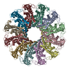
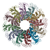
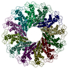
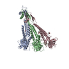
| Title | CONNECTOR PROTEIN FROM BACTERIOPHAGE PHI29 | ||||||
 Components Components | UPPER COLLAR PROTEIN | ||||||
 Keywords Keywords | VIRAL PROTEIN / helix bundle | ||||||
| Function / homology |  Function and homology information Function and homology informationviral procapsid / viral portal complex / symbiont genome ejection through host cell envelope, short tail mechanism / viral DNA genome packaging / RNA binding Similarity search - Function | ||||||
| Biological species |   Bacillus phage phi29 (virus) Bacillus phage phi29 (virus) | ||||||
| Method |  X-RAY DIFFRACTION / SYNCHROTRON / molecular replacement, MIR / Resolution: 3.2 Å X-RAY DIFFRACTION / SYNCHROTRON / molecular replacement, MIR / Resolution: 3.2 Å | ||||||
 Authors Authors | Simpson, A.A. / Leiman, P.G. / Tao, Y. / He, Y. / Badasso, M.O. / Jardine, P.J. / Anderson, D.L. / Rossmann, M.G. | ||||||
 Citation Citation | Journal: Acta Crystallogr.,Sect.D / Year: 2001 Title: Structure determination of the head-tail connector of bacteriophage phi29. Authors: Simpson, A.A. / Leiman, P.G. / Tao, Y. / He, Y. / Badasso, M.O. / Jardine, P.J. / Anderson, D.L. / Rossmann, M.G. #1: Journal: Nature / Year: 2000Title: STRUCTURE OF THE BACTERIOPHAGE PHI29 DNA PACKAGING MOTOR Authors: SIMPSON, A.A. / TAO, Y. / LEIMAN, P.G. / BADASSO, M.O. / HE, Y. / JARDINE, P.J. / OLSON, N.H. / MORAIS, M.C. / GRIMES, S. / ANDERSON, D.L. / BAKER, T.S. / ROSSMANN, M.G. | ||||||
| History |
|
- Structure visualization
| Structure viewer | Molecule: MolmilJmol/JSmol |
|---|
- Downloads & links
-Download
| PDBx/mmCIF format | 1jnb.cif.gz | 614.4 KB | Display | PDBx/mmCIF format |
|---|---|---|---|---|
| PDB format | pdb1jnb.ent.gz | 510.2 KB | Display | PDB format |
| PDBx/mmJSON format | 1jnb.json.gz | Tree view | PDBx/mmJSON format | |
| Others |  Other downloads Other downloads |
-Validation report
| Arichive directory | https://data.pdbj.org/pub/pdb/validation_reports/jn/1jnbftp://data.pdbj.org/pub/pdb/validation_reports/jn/1jnb | HTTPS FTP |
|---|
-Related structure data


-Links
 PDBj
PDBj- Assembly
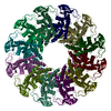
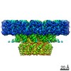
| Deposited unit | 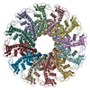
| ||||||||
|---|---|---|---|---|---|---|---|---|---|
| 1 |
| ||||||||
| Unit cell |
| ||||||||
| Details | biological unit is dodecamer, as found in crystal ASU |
-Components
| #1: Protein | Mass: 35917.293 Da / Num. of mol.: 12 Source method: isolated from a genetically manipulated source Source: (gene. exp.) Bacillus phage phi29 (virus) / Genus: Phi29-like viruses / Gene: 10 / Plasmid: PPC28D1 / Production host:  |
|---|
-Experimental details
-Experiment
| Experiment | Method: X-RAY DIFFRACTION / Number of used crystals: 1 |
|---|
- Sample preparation
| Crystal | Density Matthews: 2.95 Å3/Da / Density % sol: 57 % | ||||||||||||||||||||||||
|---|---|---|---|---|---|---|---|---|---|---|---|---|---|---|---|---|---|---|---|---|---|---|---|---|---|
| Crystal grow | Temperature: 292 K / Method: vapor diffusion, hanging drop / pH: 8 Details: 30-40% MPD, 0.1 M Tris-HCl pH 8.0, 0.05 M CaCl2, VAPOR DIFFUSION, HANGING DROP, temperature 292K | ||||||||||||||||||||||||
| Crystal grow | *PLUS Method: unknown | ||||||||||||||||||||||||
| Components of the solutions | *PLUS
|
-Data collection
| Diffraction | Mean temperature: 100 K |
|---|---|
| Diffraction source | Source: SYNCHROTRON / Site: APS  / Beamline: 14-BM-C / Wavelength: 1 Å / Beamline: 14-BM-C / Wavelength: 1 Å |
| Detector | Type: ADSC QUANTUM 4 / Detector: CCD / Date: Aug 14, 1999 |
| Radiation | Monochromator: Si 111 CHANNEL / Protocol: SINGLE WAVELENGTH / Monochromatic (M) / Laue (L): M / Scattering type: x-ray |
| Radiation wavelength | Wavelength: 1 Å / Relative weight: 1 |
| Reflection | Resolution: 3.21→50 Å / Num. all: 78427 / Num. obs: 78035 / % possible obs: 99.5 % / Observed criterion σ(F): 0 / Observed criterion σ(I): -3 / Redundancy: 3.9 % / Rmerge(I) obs: 0.032 / Net I/σ(I): 21 |
| Reflection shell | Resolution: 3.21→3.24 Å / Redundancy: 3.8 % / Rmerge(I) obs: 0.198 / Num. unique all: 2043 / % possible all: 100 |
| Reflection | *PLUS Highest resolution: 3.2 Å / Lowest resolution: 50 Å / Rmerge(I) obs: 0.065 |
| Reflection shell | *PLUS % possible obs: 99 % / Rmerge(I) obs: 0.24 |
- Processing
| Software |
| |||||||||||||||||||||||||
|---|---|---|---|---|---|---|---|---|---|---|---|---|---|---|---|---|---|---|---|---|---|---|---|---|---|---|
| Refinement | Method to determine structure: molecular replacement, MIR Starting model: low resolution model, based on cryo-EM data Resolution: 3.2→9 Å Isotropic thermal model: individual, restrained between neighbours Cross valid method: THROUGHOUT / σ(F): 3 / Stereochemistry target values: Engh & Huber / Details: CNS version 1.0 used for refinement
| |||||||||||||||||||||||||
| Solvent computation | Solvent model: solvent mask calculated using atomic coordinates | |||||||||||||||||||||||||
| Displacement parameters | Biso mean: 46.9935 Å2
| |||||||||||||||||||||||||
| Refinement step | Cycle: LAST / Resolution: 3.2→9 Å
| |||||||||||||||||||||||||
| Refine LS restraints |
| |||||||||||||||||||||||||
| Refine LS restraints NCS | NCS model details: restrained - 3 rigid domains per subunit / Rms dev position: 0.03 Å / Weight Biso : 2000 / Weight position: 300 | |||||||||||||||||||||||||
| LS refinement shell | Resolution: 3.2→3.22 Å
| |||||||||||||||||||||||||
| Software | *PLUS Name: CNS / Version: 1 / Classification: refinement | |||||||||||||||||||||||||
| Refinement | *PLUS Highest resolution: 3.2 Å / Lowest resolution: 9 Å / σ(F): 3 / % reflection Rfree: 5 % / Rfactor obs: 0.2844 | |||||||||||||||||||||||||
| Solvent computation | *PLUS | |||||||||||||||||||||||||
| Displacement parameters | *PLUS | |||||||||||||||||||||||||
| Refine LS restraints | *PLUS
| |||||||||||||||||||||||||
| LS refinement shell | *PLUS Highest resolution: 3.2 Å / % reflection Rfree: 5 % |
