 Movie
Movie Controller

[English] 日本語
 Yorodumi
- PDB-1iua: Ultra-high resolution structure of HiPIP from Thermochromatium tepidum -
+ Open data

- Basic information
| Entry | Database: PDB / ID: 1iua | |||||||||
|---|---|---|---|---|---|---|---|---|---|---|
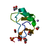
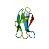
| Title | Ultra-high resolution structure of HiPIP from Thermochromatium tepidum | |||||||||
 Components Components | High-potential iron-sulfur protein | |||||||||
 Keywords Keywords | ELECTRON TRANSPORT / HiPIP | |||||||||
| Function / homology |  Function and homology information Function and homology informationaerobic electron transport chain / 4 iron, 4 sulfur cluster binding / electron transfer activity / metal ion binding Similarity search - Function | |||||||||
| Biological species |  Thermochromatium tepidum (bacteria) Thermochromatium tepidum (bacteria) | |||||||||
| Method |  X-RAY DIFFRACTION / SYNCHROTRON / MOLECULAR REPLACEMENT / Resolution: 0.8 Å X-RAY DIFFRACTION / SYNCHROTRON / MOLECULAR REPLACEMENT / Resolution: 0.8 Å | |||||||||
 Authors Authors | Liu, L. / Nogi, T. / Kobayashi, M. / Nozawa, T. / Miki, K. | |||||||||
 Citation Citation | Journal: Acta Crystallogr.,Sect.D / Year: 2002 Title: Ultrahigh-resolution structure of high-potential iron-sulfur protein from Thermochromatium tepidum. Authors: Liu, L. / Nogi, T. / Kobayashi, M. / Nozawa, T. / Miki, K. #1: Journal: Proc.Natl.Acad.Sci.USA / Year: 2000Title: CRYSTAL STRUCTURES OF PHOTOSYNTHETIC REACTION CENTER AND HIGH-POTENTIAL IRON-SULFUR PROTEIN FROM THERMOCHROMATIUM TEPIDUM: THERMOSTABILITY AND ELECTRON TRANSFER Authors: Nogi, T. / Fathir, I. / Kobayashi, M. / Nozawa, T. / Miki, K. #2: Journal: Acta Crystallogr.,Sect.D / Year: 2000Title: CRYSTALLIZATION AND PRELIMINARY CRYSTALLOGRAPHIC ANALYSIS OF HIGH-POTENTIAL IRON-SULFUR PROTEIN FROM THERMOCHROMATIUM TEPIDUM Authors: Nogi, T. / Kobayashi, M. / Nozawa, T. / Miki, K. | |||||||||
| History |
|
- Structure visualization
| Structure viewer | Molecule: MolmilJmol/JSmol |
|---|
- Downloads & links
-Download
| PDBx/mmCIF format | 1iua.cif.gz | 49.1 KB | Display | PDBx/mmCIF format |
|---|---|---|---|---|
| PDB format | pdb1iua.ent.gz | 34.4 KB | Display | PDB format |
| PDBx/mmJSON format | 1iua.json.gz | Tree view | PDBx/mmJSON format | |
| Others |  Other downloads Other downloads |
-Validation report
| Arichive directory | https://data.pdbj.org/pub/pdb/validation_reports/iu/1iuaftp://data.pdbj.org/pub/pdb/validation_reports/iu/1iua | HTTPS FTP |
|---|
-Related structure data
| Related structure data | |
|---|---|
| Similar structure data |


-Links
 PDBj
PDBj- Assembly
| Deposited unit | 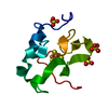
| ||||||||
|---|---|---|---|---|---|---|---|---|---|
| 1 |
| ||||||||
| Unit cell |
|
-Components
| #1: Protein | Mass: 8793.851 Da / Num. of mol.: 1 / Source method: isolated from a natural source / Source: (natural) Thermochromatium tepidum (bacteria) / References: UniProt: P80176 | ||||
|---|---|---|---|---|---|
| #2: Chemical |   Mass: 96.063 Da / Num. of mol.: 3 / Source method: obtained synthetically / Formula: SO4 Mass: 96.063 Da / Num. of mol.: 3 / Source method: obtained synthetically / Formula: SO4#3: Chemical | ChemComp-SF4 / |   Mass: 351.640 Da / Num. of mol.: 1 / Source method: obtained synthetically / Formula: Fe4S4 Mass: 351.640 Da / Num. of mol.: 1 / Source method: obtained synthetically / Formula: Fe4S4#4: Water | ChemComp-HOH / |  Mass: 18.015 Da / Num. of mol.: 93 / Source method: isolated from a natural source / Formula: H2O Mass: 18.015 Da / Num. of mol.: 93 / Source method: isolated from a natural source / Formula: H2O |
-Experimental details
-Experiment
| Experiment | Method: X-RAY DIFFRACTION / Number of used crystals: 1 |
|---|
- Sample preparation
| Crystal | Density Matthews: 1.74 Å3/Da / Density % sol: 29.17 % | ||||||||||||||||||||||||
|---|---|---|---|---|---|---|---|---|---|---|---|---|---|---|---|---|---|---|---|---|---|---|---|---|---|
| Crystal grow | Temperature: 293.2 K / Method: vapor diffusion, sitting drop / pH: 3.5 Details: 0.7M A/S, 0.05M Sodium Citrate, pH 3.5, VAPOR DIFFUSION, SITTING DROP, temperature 293.2K | ||||||||||||||||||||||||
| Crystal grow | *PLUS Temperature: 293 K / pH: 8 | ||||||||||||||||||||||||
| Components of the solutions | *PLUS
|
-Data collection
| Diffraction | Mean temperature: 100 K |
|---|---|
| Diffraction source | Source: SYNCHROTRON / Site: SPring-8  / Beamline: BL40B2 / Wavelength: 0.7293 Å / Beamline: BL40B2 / Wavelength: 0.7293 Å |
| Detector | Type: RIGAKU RAXIS IV / Detector: IMAGE PLATE / Date: Mar 25, 2000 |
| Radiation | Monochromator: undulator / Protocol: SINGLE WAVELENGTH / Monochromatic (M) / Laue (L): M / Scattering type: x-ray |
| Radiation wavelength | Wavelength: 0.7293 Å / Relative weight: 1 |
| Reflection | Resolution: 0.8→50 Å / Num. all: 65310 / Num. obs: 64373 / % possible obs: 98.6 % / Observed criterion σ(F): 2 / Observed criterion σ(I): 2 |
| Reflection shell | Resolution: 0.8→0.81 Å / % possible all: 95.5 |
| Reflection | *PLUS Lowest resolution: 5 Å / Num. measured all: 823255 / Rmerge(I) obs: 0.052 |
| Reflection shell | *PLUS % possible obs: 95.5 % / Num. unique obs: 3094 / Rmerge(I) obs: 0.397 / Mean I/σ(I) obs: 2.74 |
- Processing
| Software |
| ||||||||||||||||
|---|---|---|---|---|---|---|---|---|---|---|---|---|---|---|---|---|---|
| Refinement | Method to determine structure: MOLECULAR REPLACEMENT / Resolution: 0.8→20 Å / σ(F): 4 / Stereochemistry target values: Engh & Huber / Details: No restraint for the last full-matrix refinement
| ||||||||||||||||
| Refinement step | Cycle: LAST / Resolution: 0.8→20 Å
| ||||||||||||||||
| Refine LS restraints |
| ||||||||||||||||
| Software | *PLUS Name: SHELXL / Version: 97 / Classification: refinement | ||||||||||||||||
| Refinement | *PLUS Lowest resolution: 20 Å / Rfactor Rwork: 0.101 | ||||||||||||||||
| Solvent computation | *PLUS | ||||||||||||||||
| Displacement parameters | *PLUS | ||||||||||||||||
| Refine LS restraints | *PLUS
|
