 Movie
Movie Controller


+ Open data

- Basic information
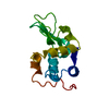
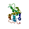
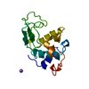
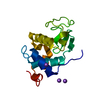
| Entry | Database: PDB / ID: 1ioc | ||||||
|---|---|---|---|---|---|---|---|
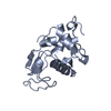
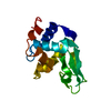
| Title | CRYSTAL STRUCTURE OF MUTANT HUMAN LYSOZYME, EAEA-I56T | ||||||
 Components Components | LYSOZYME C | ||||||
 Keywords Keywords | HYDROLASE / amyloid / mutant / human lysozyme / stability | ||||||
| Function / homology |  Function and homology information Function and homology informationantimicrobial humoral response / Antimicrobial peptides / specific granule lumen / azurophil granule lumen / tertiary granule lumen / lysozyme / lysozyme activity / defense response to Gram-negative bacterium / killing of cells of another organism / defense response to Gram-positive bacterium ...antimicrobial humoral response / Antimicrobial peptides / specific granule lumen / azurophil granule lumen / tertiary granule lumen / lysozyme / lysozyme activity / defense response to Gram-negative bacterium / killing of cells of another organism / defense response to Gram-positive bacterium / defense response to bacterium / inflammatory response / Amyloid fiber formation / Neutrophil degranulation / extracellular space / extracellular exosome / extracellular region / identical protein binding Similarity search - Function | ||||||
| Biological species |  Homo sapiens (human) Homo sapiens (human) | ||||||
| Method |  X-RAY DIFFRACTION / SYNCHROTRON / MOLECULAR REPLACEMENT / Resolution: 2.4 Å X-RAY DIFFRACTION / SYNCHROTRON / MOLECULAR REPLACEMENT / Resolution: 2.4 Å | ||||||
 Authors Authors | Goda, S. / Takano, K. / Yamagata, Y. / Yutani, K. | ||||||
 Citation Citation | Journal: J.Biochem. / Year: 2002 Title: Elongation in a beta-structure promotes amyloid-like fibril formation of human lysozyme. Authors: Goda, S. / Takano, K. / Yamagata, Y. / Maki, S. / Namba, K. / Yutani, K. #1: Journal: Protein Eng. / Year: 2000Title: Effect of extra N-terminal residues on the stability and folding of human lysozyme expressed in Pichia pastoris Authors: Goda, S. / Takano, K. / Yamagata, Y. / Katakura, Y. / Yutani, K. | ||||||
| History |
|
- Structure visualization
| Structure viewer | Molecule: MolmilJmol/JSmol |
|---|
- Downloads & links
-Download
| PDBx/mmCIF format | 1ioc.cif.gz | 40.8 KB | Display | PDBx/mmCIF format |
|---|---|---|---|---|
| PDB format | pdb1ioc.ent.gz | 28.4 KB | Display | PDB format |
| PDBx/mmJSON format | 1ioc.json.gz | Tree view | PDBx/mmJSON format | |
| Others |  Other downloads Other downloads |
-Validation report
| Summary document | 1ioc_validation.pdf.gz | 371.2 KB | Display | wwPDB validaton report |
|---|---|---|---|---|
| Full document | 1ioc_full_validation.pdf.gz | 374.2 KB | Display | |
| Data in XML | 1ioc_validation.xml.gz | 4.5 KB | Display | |
| Data in CIF | 1ioc_validation.cif.gz | 6.8 KB | Display | |
| Arichive directory | https://data.pdbj.org/pub/pdb/validation_reports/io/1iocftp://data.pdbj.org/pub/pdb/validation_reports/io/1ioc | HTTPS FTP |
-Related structure data
| Related structure data | |
|---|---|
| Similar structure data |


-Links
 PDBj
PDBj


- Assembly
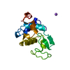
| Deposited unit | 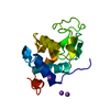
| ||||||||
|---|---|---|---|---|---|---|---|---|---|
| 1 |
| ||||||||
| Unit cell |
|
-Components
| #1: Protein | Mass: 15109.023 Da / Num. of mol.: 1 / Mutation: I56T Source method: isolated from a genetically manipulated source Source: (gene. exp.) Homo sapiens (human) / Plasmid: PPIC9 / Production host:  Pichia pastoris (fungus) / References: UniProt: P61626, lysozyme Pichia pastoris (fungus) / References: UniProt: P61626, lysozyme | ||
|---|---|---|---|
| #2: Chemical |   Mass: 22.990 Da / Num. of mol.: 3 / Source method: obtained synthetically / Formula: Na Mass: 22.990 Da / Num. of mol.: 3 / Source method: obtained synthetically / Formula: Na#3: Water | ChemComp-HOH / |  Mass: 18.015 Da / Num. of mol.: 88 / Source method: isolated from a natural source / Formula: H2O Mass: 18.015 Da / Num. of mol.: 88 / Source method: isolated from a natural source / Formula: H2O |
-Experimental details
-Experiment
| Experiment | Method: X-RAY DIFFRACTION / Number of used crystals: 1 |
|---|
- Sample preparation
| Crystal | Density Matthews: 3.59 Å3/Da / Density % sol: 65.77 % | |||||||||||||||||||||||||
|---|---|---|---|---|---|---|---|---|---|---|---|---|---|---|---|---|---|---|---|---|---|---|---|---|---|---|
| Crystal grow | Temperature: 283 K / Method: vapor diffusion, hanging drop / pH: 4.6 Details: cadmium, chloride, sodium acetate, PEG400, pH 4.6, VAPOR DIFFUSION, HANGING DROP, temperature 283K | |||||||||||||||||||||||||
| Crystal grow | *PLUS Temperature: 10 ℃ / Details: Goda, S., (2000) Protein Eng., 13, 299. | |||||||||||||||||||||||||
| Components of the solutions | *PLUS
|
-Data collection
| Diffraction | Mean temperature: 100 K |
|---|---|
| Diffraction source | Source: SYNCHROTRON / Site: Photon Factory  / Beamline: BL-18B / Wavelength: 1 Å / Beamline: BL-18B / Wavelength: 1 Å |
| Detector | Type: WEISSENBERG / Detector: DIFFRACTOMETER / Date: Nov 27, 1999 |
| Radiation | Protocol: SINGLE WAVELENGTH / Monochromatic (M) / Laue (L): M / Scattering type: x-ray |
| Radiation wavelength | Wavelength: 1 Å / Relative weight: 1 |
| Reflection | Highest resolution: 2.4 Å / Num. all: 72930 / Num. obs: 9168 / % possible obs: 99.8 % / Rmerge(I) obs: 0.118 |
| Reflection | *PLUS Num. measured all: 72930 |
- Processing
| Software |
| ||||||||||||
|---|---|---|---|---|---|---|---|---|---|---|---|---|---|
| Refinement | Method to determine structure: MOLECULAR REPLACEMENT Starting model: EAEA human lysozyme Resolution: 2.4→6 Å
| ||||||||||||
| Refinement step | Cycle: LAST / Resolution: 2.4→6 Å
|
