 Movie
Movie Controller

[English] 日本語
 Yorodumi
- PDB-1i7d: NONCOVALENT COMPLEX OF E.COLI DNA TOPOISOMERASE III WITH AN 8-BAS... -
+ Open data

- Basic information
| Entry | Database: PDB / ID: 1i7d | ||||||
|---|---|---|---|---|---|---|---|
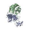
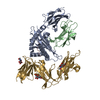
| Title | NONCOVALENT COMPLEX OF E.COLI DNA TOPOISOMERASE III WITH AN 8-BASE SINGLE-STRANDED DNA OLIGONUCLEOTIDE | ||||||
 Components Components |
| ||||||
 Keywords Keywords | ISOMERASE/DNA / DNA topoisomerase / decatenating enzyme / protein-DNA complex / single-stranded DNA / ISOMERASE-DNA COMPLEX | ||||||
| Function / homology |  Function and homology information Function and homology informationcytoplasmic replication fork / sequence-specific single stranded DNA binding / chromosome separation / DNA topoisomerase / DNA topoisomerase type I (single strand cut, ATP-independent) activity / DNA topological change / DNA-templated DNA replication / DNA recombination / DNA repair / magnesium ion binding Similarity search - Function | ||||||
| Biological species |  | ||||||
| Method |  X-RAY DIFFRACTION / SYNCHROTRON / MOLECULAR REPLACEMENT / Resolution: 2.05 Å X-RAY DIFFRACTION / SYNCHROTRON / MOLECULAR REPLACEMENT / Resolution: 2.05 Å | ||||||
 Authors Authors | Changela, A. / DiGate, R.J. / Mondragon, A. | ||||||
 Citation Citation | Journal: Nature / Year: 2001 Title: Crystal structure of a complex of a type IA DNA topoisomerase with a single-stranded DNA molecule. Authors: Changela, A. / DiGate, R.J. / Mondragon, A. | ||||||
| History |
|
- Structure visualization
| Structure viewer | Molecule: MolmilJmol/JSmol |
|---|
- Downloads & links
-Download
| PDBx/mmCIF format | 1i7d.cif.gz | 148.7 KB | Display | PDBx/mmCIF format |
|---|---|---|---|---|
| PDB format | pdb1i7d.ent.gz | 113.3 KB | Display | PDB format |
| PDBx/mmJSON format | 1i7d.json.gz | Tree view | PDBx/mmJSON format | |
| Others |  Other downloads Other downloads |
-Validation report
| Arichive directory | https://data.pdbj.org/pub/pdb/validation_reports/i7/1i7dftp://data.pdbj.org/pub/pdb/validation_reports/i7/1i7d | HTTPS FTP |
|---|
-Related structure data
| Related structure data |  1d6mS S: Starting model for refinement |
|---|---|
| Similar structure data |


-Links
 PDBj
PDBj


- Assembly
| Deposited unit | 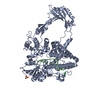
| ||||||||
|---|---|---|---|---|---|---|---|---|---|
| 1 |
| ||||||||
| Unit cell |
|
-Components
| #1: DNA chain | Mass: 2386.593 Da / Num. of mol.: 1 / Source method: obtained synthetically |
|---|---|
| #2: Protein | Mass: 74135.680 Da / Num. of mol.: 1 / Mutation: Y328F Source method: isolated from a genetically manipulated source Source: (gene. exp.) |
| #3: Chemical | ChemComp-CL /   Mass: 35.453 Da / Num. of mol.: 1 / Source method: obtained synthetically / Formula: Cl Mass: 35.453 Da / Num. of mol.: 1 / Source method: obtained synthetically / Formula: Cl |
| #4: Chemical | ChemComp-SO4 /   Mass: 96.063 Da / Num. of mol.: 1 / Source method: obtained synthetically / Formula: SO4 Mass: 96.063 Da / Num. of mol.: 1 / Source method: obtained synthetically / Formula: SO4 |
| #5: Water | ChemComp-HOH /  Mass: 18.015 Da / Num. of mol.: 332 / Source method: isolated from a natural source / Formula: H2O Mass: 18.015 Da / Num. of mol.: 332 / Source method: isolated from a natural source / Formula: H2O |
-Experimental details
-Experiment
| Experiment | Method: X-RAY DIFFRACTION / Number of used crystals: 1 |
|---|
- Sample preparation
| Crystal | Density Matthews: 3.03 Å3/Da / Density % sol: 59.46 % | ||||||||||||||||||||||||||||||
|---|---|---|---|---|---|---|---|---|---|---|---|---|---|---|---|---|---|---|---|---|---|---|---|---|---|---|---|---|---|---|---|
| Crystal grow | Temperature: 295 K / Method: vapor diffusion, hanging drop / pH: 5.5 Details: 1.3M ammonium sulfate, 0.8M sodium chloride, 0.1M sodium citrate, pH 5.5, VAPOR DIFFUSION, HANGING DROP, temperature 295K | ||||||||||||||||||||||||||||||
| Components of the solutions |
| ||||||||||||||||||||||||||||||
| Crystal grow | *PLUS Temperature: 22 ℃ | ||||||||||||||||||||||||||||||
| Components of the solutions | *PLUS
|
-Data collection
| Diffraction | Mean temperature: 100 K |
|---|---|
| Diffraction source | Source: SYNCHROTRON / Site: APS  / Beamline: 5ID-B / Wavelength: 0.9948 Å / Beamline: 5ID-B / Wavelength: 0.9948 Å |
| Detector | Type: MARRESEARCH / Detector: CCD / Date: Jun 1, 2000 |
| Radiation | Protocol: SINGLE WAVELENGTH / Monochromatic (M) / Laue (L): M / Scattering type: x-ray |
| Radiation wavelength | Wavelength: 0.9948 Å / Relative weight: 1 |
| Reflection | Resolution: 2.05→29.63 Å / Num. all: 296621 / Num. obs: 55797 / % possible obs: 96.2 % / Observed criterion σ(F): 0 / Observed criterion σ(I): 0 / Redundancy: 5.3 % / Biso Wilson estimate: 25.7 Å2 / Rmerge(I) obs: 0.098 / Rsym value: 9 / Net I/σ(I): 5.5 |
| Reflection shell | Resolution: 2.05→2.1 Å / Redundancy: 3.5 % / Rmerge(I) obs: 0.244 / Mean I/σ(I) obs: 2.7 / Num. unique all: 4129 / Rsym value: 20.7 / % possible all: 96.9 |
| Reflection | *PLUS Num. measured all: 296621 |
| Reflection shell | *PLUS % possible obs: 96.9 % |
- Processing
| Software |
| ||||||||||||||||||||||||||||||||||||||||
|---|---|---|---|---|---|---|---|---|---|---|---|---|---|---|---|---|---|---|---|---|---|---|---|---|---|---|---|---|---|---|---|---|---|---|---|---|---|---|---|---|---|
| Refinement | Method to determine structure: MOLECULAR REPLACEMENT Starting model: PDB entry 1D6M Resolution: 2.05→29.63 Å / Rfactor Rfree error: 0.005 / Data cutoff high absF: 1962525.77 / Data cutoff low absF: 0 / Isotropic thermal model: RESTRAINED / Cross valid method: THROUGHOUT / σ(F): 0 / σ(I): 0 / Stereochemistry target values: Engh & Huber / Details: Refinement done in CNS 1.0
| ||||||||||||||||||||||||||||||||||||||||
| Solvent computation | Solvent model: FLAT MODEL / Bsol: 53.73 Å2 / ksol: 0.39 e/Å3 | ||||||||||||||||||||||||||||||||||||||||
| Displacement parameters | Biso mean: 47.8 Å2
| ||||||||||||||||||||||||||||||||||||||||
| Refine analyze |
| ||||||||||||||||||||||||||||||||||||||||
| Refinement step | Cycle: LAST / Resolution: 2.05→29.63 Å
| ||||||||||||||||||||||||||||||||||||||||
| Refine LS restraints |
| ||||||||||||||||||||||||||||||||||||||||
| LS refinement shell | Resolution: 2.05→2.18 Å / Rfactor Rfree error: 0.014 / Total num. of bins used: 6
| ||||||||||||||||||||||||||||||||||||||||
| Xplor file |
| ||||||||||||||||||||||||||||||||||||||||
| Software | *PLUS Name: CNS / Version: 1 / Classification: refinement | ||||||||||||||||||||||||||||||||||||||||
| Refinement | *PLUS σ(F): 0 / % reflection Rfree: 5.1 % / Rfactor Rfree: 0.26 | ||||||||||||||||||||||||||||||||||||||||
| Solvent computation | *PLUS | ||||||||||||||||||||||||||||||||||||||||
| Displacement parameters | *PLUS Biso mean: 47.8 Å2 | ||||||||||||||||||||||||||||||||||||||||
| Refine LS restraints | *PLUS
| ||||||||||||||||||||||||||||||||||||||||
| LS refinement shell | *PLUS Rfactor Rfree: 0.304 / % reflection Rfree: 5.1 % / Rfactor Rwork: 0.301 |
