 Movie
Movie Controller

[English] 日本語
 Yorodumi
- PDB-2o59: Structure of E. coli topoisomerase III in complex with an 8-base ... -
+ Open data

- Basic information
| Entry | Database: PDB / ID: 2o59 | ||||||
|---|---|---|---|---|---|---|---|
| Title | Structure of E. coli topoisomerase III in complex with an 8-base single stranded oligonucleotide. Frozen in glycerol pH 8.0 | ||||||
 Components Components |
| ||||||
 Keywords Keywords | ISOMERASE/DNA / topoisomerase type IA complex with ssDNA / ISOMERASE-DNA COMPLEX | ||||||
| Function / homology |  Function and homology information Function and homology informationcytoplasmic replication fork / sequence-specific single stranded DNA binding / chromosome separation / DNA topoisomerase / DNA topoisomerase type I (single strand cut, ATP-independent) activity / DNA topological change / DNA-templated DNA replication / DNA recombination / DNA repair / magnesium ion binding Similarity search - Function | ||||||
| Biological species |  | ||||||
| Method |  X-RAY DIFFRACTION / SYNCHROTRON / MOLECULAR REPLACEMENT / Resolution: 2.5 Å X-RAY DIFFRACTION / SYNCHROTRON / MOLECULAR REPLACEMENT / Resolution: 2.5 Å | ||||||
 Authors Authors | Changela, A. / DiGate, R.J. / Mondragon, A. | ||||||
 Citation Citation | Journal: J.Mol.Biol. / Year: 2007 Title: Structural Studies of E. coli Topoisomerase III-DNA Complexes Reveal a Novel Type IA Topoisomerase-DNA Conformational Intermediate. Authors: Changela, A. / Digate, R.J. / Mondragon, A. | ||||||
| History |
|
- Structure visualization
| Structure viewer | Molecule: MolmilJmol/JSmol |
|---|
- Downloads & links
-Download
| PDBx/mmCIF format | 2o59.cif.gz | 511 KB | Display | PDBx/mmCIF format |
|---|---|---|---|---|
| PDB format | pdb2o59.ent.gz | 424.5 KB | Display | PDB format |
| PDBx/mmJSON format | 2o59.json.gz | Tree view | PDBx/mmJSON format | |
| Others |  Other downloads Other downloads |
-Validation report
| Arichive directory | https://data.pdbj.org/pub/pdb/validation_reports/o5/2o59ftp://data.pdbj.org/pub/pdb/validation_reports/o5/2o59 | HTTPS FTP |
|---|
-Related structure data
| Related structure data |  2o19C  2o54C  2o5cC  2o5eC  1d6mS  1i7dS S: Starting model for refinement C: citing same article ( |
|---|---|
| Similar structure data |


-Links
 PDBj
PDBj


- Assembly
| Deposited unit | 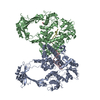
| ||||||||
|---|---|---|---|---|---|---|---|---|---|
| 1 | 
| ||||||||
| 2 | 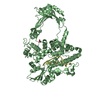
| ||||||||
| Unit cell |
| ||||||||
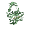
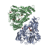
| Details | Complex of E. coli topoisomerase III with DNA. Two molecules in the asymmetric unit. Each molecule in a different conformational state. |
-Components
| #1: DNA chain | Mass: 2386.593 Da / Num. of mol.: 2 / Source method: obtained synthetically #2: Protein | Mass: 74151.680 Da / Num. of mol.: 2 Source method: isolated from a genetically manipulated source Source: (gene. exp.) #3: Chemical |   Mass: 35.453 Da / Num. of mol.: 2 / Source method: obtained synthetically / Formula: Cl Mass: 35.453 Da / Num. of mol.: 2 / Source method: obtained synthetically / Formula: Cl#4: Chemical | ChemComp-ACY / |   Mass: 60.052 Da / Num. of mol.: 1 / Source method: obtained synthetically / Formula: C2H4O2 Mass: 60.052 Da / Num. of mol.: 1 / Source method: obtained synthetically / Formula: C2H4O2#5: Water | ChemComp-HOH / |  Mass: 18.015 Da / Num. of mol.: 142 / Source method: isolated from a natural source / Formula: H2O Mass: 18.015 Da / Num. of mol.: 142 / Source method: isolated from a natural source / Formula: H2O |
|---|
-Experimental details
-Experiment
| Experiment | Method: X-RAY DIFFRACTION / Number of used crystals: 1 |
|---|
- Sample preparation
| Crystal | Density Matthews: 3.82 Å3/Da / Density % sol: 67.77 % | ||||||||||||||||||||||||||||||||||||
|---|---|---|---|---|---|---|---|---|---|---|---|---|---|---|---|---|---|---|---|---|---|---|---|---|---|---|---|---|---|---|---|---|---|---|---|---|---|
| Crystal grow | Temperature: 295 K / Method: vapor diffusion, hanging drop / pH: 8 Details: 1.5 M (NH4)SO4, 0.1 M Sodium citrate, 0.5 M NaCl, pH 8.0, VAPOR DIFFUSION, HANGING DROP, temperature 295K | ||||||||||||||||||||||||||||||||||||
| Components of the solutions |
|
-Data collection
| Diffraction | Mean temperature: 100 K |
|---|---|
| Diffraction source | Source: SYNCHROTRON / Site: APS  / Beamline: 5ID-B / Wavelength: 1 Å / Beamline: 5ID-B / Wavelength: 1 Å |
| Detector | Type: MARMOSAIC 225 mm CCD / Detector: CCD / Date: Jan 15, 2003 |
| Radiation | Protocol: SINGLE WAVELENGTH / Monochromatic (M) / Laue (L): M / Scattering type: x-ray |
| Radiation wavelength | Wavelength: 1 Å / Relative weight: 1 |
| Reflection | Resolution: 2.503→29.534 Å / Num. obs: 81050 / % possible obs: 97.8 % / Redundancy: 5.2 % / Rmerge(I) obs: 0.05 / Rsym value: 0.05 / Net I/σ(I): 10.8 |
| Reflection shell | Resolution: 2.5→2.56 Å / Redundancy: 3 % / Rmerge(I) obs: 0.269 / Mean I/σ(I) obs: 2.8 / Num. measured all: 15495 / Num. unique all: 5081 / Rsym value: 0.269 / % possible all: 88.2 |
- Processing
| Software |
| ||||||||||||||||||||||||||||||||||||||||||||||||||||||||||||||||||||||||||||||||||||||||||
|---|---|---|---|---|---|---|---|---|---|---|---|---|---|---|---|---|---|---|---|---|---|---|---|---|---|---|---|---|---|---|---|---|---|---|---|---|---|---|---|---|---|---|---|---|---|---|---|---|---|---|---|---|---|---|---|---|---|---|---|---|---|---|---|---|---|---|---|---|---|---|---|---|---|---|---|---|---|---|---|---|---|---|---|---|---|---|---|---|---|---|---|
| Refinement | Method to determine structure: MOLECULAR REPLACEMENT Starting model: PDB ENTRY 1D6M, 1I7D Resolution: 2.5→29.534 Å / Cor.coef. Fo:Fc: 0.93 / Cor.coef. Fo:Fc free: 0.909 / SU B: 14.55 / SU ML: 0.176 / Cross valid method: THROUGHOUT / σ(F): 0 / ESU R: 0.306 / ESU R Free: 0.243 / Stereochemistry target values: MAXIMUM LIKELIHOOD / Details: HYDROGENS HAVE BEEN ADDED IN THE RIDING POSITIONS
| ||||||||||||||||||||||||||||||||||||||||||||||||||||||||||||||||||||||||||||||||||||||||||
| Solvent computation | Ion probe radii: 0.8 Å / Shrinkage radii: 0.8 Å / VDW probe radii: 1.4 Å / Solvent model: MASK | ||||||||||||||||||||||||||||||||||||||||||||||||||||||||||||||||||||||||||||||||||||||||||
| Displacement parameters | Biso mean: 45.095 Å2
| ||||||||||||||||||||||||||||||||||||||||||||||||||||||||||||||||||||||||||||||||||||||||||
| Refinement step | Cycle: LAST / Resolution: 2.5→29.534 Å
| ||||||||||||||||||||||||||||||||||||||||||||||||||||||||||||||||||||||||||||||||||||||||||
| Refine LS restraints |
| ||||||||||||||||||||||||||||||||||||||||||||||||||||||||||||||||||||||||||||||||||||||||||
| LS refinement shell | Resolution: 2.503→2.568 Å / Total num. of bins used: 20
|
