 Movie
Movie Controller

[English] 日本語
 Yorodumi
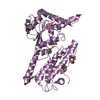
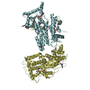
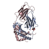
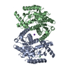
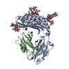
- PDB-1hw7: HSP33, HEAT SHOCK PROTEIN WITH REDOX-REGULATED CHAPERONE ACTIVITY -
+ Open data

- Basic information
| Entry | Database: PDB / ID: 1hw7 | ||||||
|---|---|---|---|---|---|---|---|
| Title | HSP33, HEAT SHOCK PROTEIN WITH REDOX-REGULATED CHAPERONE ACTIVITY | ||||||
 Components Components | HEAT SHOCK PROTEIN HSP33 | ||||||
 Keywords Keywords | CHAPERONE / Hsp33 / dimerization / heat shock protein / oxidative stress | ||||||
| Function / homology |  Function and homology information Function and homology informationmaintenance of unfolded protein / protein folding chaperone / unfolded protein binding / response to heat / protein refolding / response to oxidative stress / zinc ion binding / identical protein binding / cytoplasm / cytosol Similarity search - Function | ||||||
| Biological species |  | ||||||
| Method |  X-RAY DIFFRACTION / SYNCHROTRON / MAD / Resolution: 2.2 Å X-RAY DIFFRACTION / SYNCHROTRON / MAD / Resolution: 2.2 Å | ||||||
 Authors Authors | Vijayalakshmi, J. / Mukhergee, M.K. / Graumann, J. / Jakob, U. / Saper, M.A. | ||||||
 Citation Citation | Journal: Structure / Year: 2001 Title: The 2.2 A crystal structure of Hsp33: a heat shock protein with redox-regulated chaperone activity. Authors: Vijayalakshmi, J. / Mukhergee, M.K. / Graumann, J. / Jakob, U. / Saper, M.A. | ||||||
| History |
|
- Structure visualization
| Structure viewer | Molecule: MolmilJmol/JSmol |
|---|
- Downloads & links
-Download
| PDBx/mmCIF format | 1hw7.cif.gz | 63.5 KB | Display | PDBx/mmCIF format |
|---|---|---|---|---|
| PDB format | pdb1hw7.ent.gz | 46.2 KB | Display | PDB format |
| PDBx/mmJSON format | 1hw7.json.gz | Tree view | PDBx/mmJSON format | |
| Others |  Other downloads Other downloads |
-Validation report
| Arichive directory | https://data.pdbj.org/pub/pdb/validation_reports/hw/1hw7ftp://data.pdbj.org/pub/pdb/validation_reports/hw/1hw7 | HTTPS FTP |
|---|
-Related structure data
| Similar structure data |
|---|


-Links
 PDBj
PDBj
- Assembly
| Deposited unit | 
| ||||||||
|---|---|---|---|---|---|---|---|---|---|
| 1 | 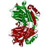
| ||||||||
| Unit cell |
| ||||||||
| Details | The second part of the biological assembly is generated by the two fold axis: -Y, -X, -Z+1/6 applied to chain A |
-Components
| #1: Protein | Mass: 28442.018 Da / Num. of mol.: 1 / Fragment: N-TERMINAL DOMAIN Source method: isolated from a genetically manipulated source Source: (gene. exp.) | ||||||
|---|---|---|---|---|---|---|---|
| #2: Chemical | ChemComp-SO4 /   Mass: 96.063 Da / Num. of mol.: 4 / Source method: obtained synthetically / Formula: SO4 Mass: 96.063 Da / Num. of mol.: 4 / Source method: obtained synthetically / Formula: SO4#3: Chemical | ChemComp-ZN / |   Mass: 65.409 Da / Num. of mol.: 1 / Source method: obtained synthetically / Formula: Zn Mass: 65.409 Da / Num. of mol.: 1 / Source method: obtained synthetically / Formula: Zn#4: Chemical | ChemComp-MES / |   Mass: 195.237 Da / Num. of mol.: 1 / Source method: obtained synthetically / Formula: C6H13NO4S / Comment: pH buffer*YM Mass: 195.237 Da / Num. of mol.: 1 / Source method: obtained synthetically / Formula: C6H13NO4S / Comment: pH buffer*YM#5: Water | ChemComp-HOH / |  Mass: 18.015 Da / Num. of mol.: 144 / Source method: isolated from a natural source / Formula: H2O Mass: 18.015 Da / Num. of mol.: 144 / Source method: isolated from a natural source / Formula: H2O |
-Experimental details
-Experiment
| Experiment | Method: X-RAY DIFFRACTION / Number of used crystals: 1 |
|---|
- Sample preparation
| Crystal | Density Matthews: 3.12 Å3/Da / Density % sol: 60.6 % | ||||||||||||||||||||||||||||||||||||||||||||||||
|---|---|---|---|---|---|---|---|---|---|---|---|---|---|---|---|---|---|---|---|---|---|---|---|---|---|---|---|---|---|---|---|---|---|---|---|---|---|---|---|---|---|---|---|---|---|---|---|---|---|
| Crystal grow | Temperature: 298 K / Method: vapor diffusion, hanging drop / pH: 6.8 Details: Drops contained 2 microlitres of protein solution (10mg/ml in 20mM KCL, 40mM HEPES pH 7.5) supplemented with 2mM DTT and 0.3 mM ZnCl2 and 2 microlitres of precipitant (1.5 M ammonium ...Details: Drops contained 2 microlitres of protein solution (10mg/ml in 20mM KCL, 40mM HEPES pH 7.5) supplemented with 2mM DTT and 0.3 mM ZnCl2 and 2 microlitres of precipitant (1.5 M ammonium sulfate, 10% dioxane, 0.1 M MES pH 6.8) and were equilibrated against 1ml of the same precipitant., VAPOR DIFFUSION, HANGING DROP, temperature 298K | ||||||||||||||||||||||||||||||||||||||||||||||||
| Crystal grow | *PLUS Temperature: 22 ℃ / pH: 7.5 | ||||||||||||||||||||||||||||||||||||||||||||||||
| Components of the solutions | *PLUS
|
-Data collection
| Diffraction | Mean temperature: 100 K |
|---|---|
| Diffraction source | Source: SYNCHROTRON / Site: APS  / Beamline: 14-BM-C / Wavelength: 1 Å / Beamline: 14-BM-C / Wavelength: 1 Å |
| Detector | Type: ADSC QUANTUM 4 / Detector: CCD / Date: Mar 31, 2000 / Details: Bent conical Si-mirror (Rh coating) |
| Radiation | Monochromator: Bent cylindrical Ge(111) monochromator / Protocol: MAD / Monochromatic (M) / Laue (L): M / Scattering type: x-ray |
| Radiation wavelength | Wavelength: 1 Å / Relative weight: 1 |
| Reflection | Resolution: 2.2→30 Å / Num. all: 18629 / Num. obs: 18629 / % possible obs: 99.7 % / Observed criterion σ(F): 0 / Observed criterion σ(I): 0 / Redundancy: 23.9 % / Biso Wilson estimate: 32.2 Å2 / Rmerge(I) obs: 0.048 / Net I/σ(I): 45.7 |
| Reflection shell | Resolution: 2.2→2.28 Å / Redundancy: 6.9 % / Rmerge(I) obs: 0.323 / Mean I/σ(I) obs: 14 / Num. unique all: 1968 / % possible all: 99.9 |
| Reflection | *PLUS Num. measured all: 444763 |
| Reflection shell | *PLUS % possible obs: 99.9 % |
- Processing
| Software |
| ||||||||||||||||||||||||||||||||||||||||
|---|---|---|---|---|---|---|---|---|---|---|---|---|---|---|---|---|---|---|---|---|---|---|---|---|---|---|---|---|---|---|---|---|---|---|---|---|---|---|---|---|---|
| Refinement | Method to determine structure: MAD Starting model: Structure phased by three-wavelength MAD experiment of AUCN-derived crystals and refined to 2.4 A. Resolution: 2.2→30 Å / Cross valid method: Free R / σ(F): 0 / σ(I): 0 / Stereochemistry target values: Engh & Huber Details: Used Bijovoet pairs of reflections, anomalous corrections to scattering factors for Zn at 1A and bulk solvent corrections
| ||||||||||||||||||||||||||||||||||||||||
| Solvent computation | Bsol: 77.4 Å2 / ksol: 0.38 e/Å3 | ||||||||||||||||||||||||||||||||||||||||
| Displacement parameters | Biso mean: 39.9 Å2 | ||||||||||||||||||||||||||||||||||||||||
| Refine analyze |
| ||||||||||||||||||||||||||||||||||||||||
| Refinement step | Cycle: LAST / Resolution: 2.2→30 Å
| ||||||||||||||||||||||||||||||||||||||||
| Refine LS restraints |
| ||||||||||||||||||||||||||||||||||||||||
| Software | *PLUS Name: CNS / Version: 0.9 / Classification: refinement | ||||||||||||||||||||||||||||||||||||||||
| Refinement | *PLUS Highest resolution: 2.2 Å / Lowest resolution: 30 Å / σ(F): 0 / % reflection Rfree: 5 % / Rfactor obs: 0.229 | ||||||||||||||||||||||||||||||||||||||||
| Solvent computation | *PLUS | ||||||||||||||||||||||||||||||||||||||||
| Displacement parameters | *PLUS Biso mean: 39.9 Å2 | ||||||||||||||||||||||||||||||||||||||||
| Refine LS restraints | *PLUS
|
