 Movie
Movie Controller

[English] 日本語
 Yorodumi
- PDB-1hjs: Structure of two fungal beta-1,4-galactanases: searching for the ... -
+ Open data

- Basic information
| Entry | Database: PDB / ID: 1hjs | ||||||
|---|---|---|---|---|---|---|---|
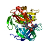
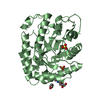
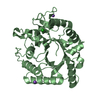
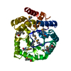
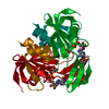
| Title | Structure of two fungal beta-1,4-galactanases: searching for the basis for temperature and pH optimum. | ||||||
 Components Components | BETA-1,4-GALACTANASE | ||||||
 Keywords Keywords | HYDROLASE / BETA-1 / 4-GALACTANASES / FAMILY 53 GLYCOSIDE HYDROLASE / THERMOSTABILITY / PH OPTIMUM / CLAN GH-A / THERMOPHILE / ALKALOPHILE | ||||||
| Function / homology |  Function and homology information Function and homology informationarabinogalactan endo-beta-1,4-galactanase / arabinogalactan endo-1,4-beta-galactosidase activity / glucosidase activity / pectin catabolic process / polysaccharide binding / hydrolase activity, hydrolyzing O-glycosyl compounds / cell wall macromolecule catabolic process Similarity search - Function | ||||||
| Biological species |  THIELAVIA HETEROTHALLICA (fungus) THIELAVIA HETEROTHALLICA (fungus) | ||||||
| Method |  X-RAY DIFFRACTION / SYNCHROTRON / OTHER / Resolution: 1.87 Å X-RAY DIFFRACTION / SYNCHROTRON / OTHER / Resolution: 1.87 Å | ||||||
 Authors Authors | Le Nours, J. / Ryttersgaard, C. / Lo Leggio, L. / Ostergaard, P.R. / Borchert, T.V. / Christensen, L.L.H. / Larsen, S. | ||||||
 Citation Citation | Journal: Protein Sci. / Year: 2003 Title: Structure of Two Fungal Beta-1,4-Galactanases: Searching for the Basis for Temperature and Ph Optimum Authors: Le Nours, J. / Ryttersgaard, C. / Lo Leggio, L. / Ostergaard, P.R. / Borchert, T.V. / Christensen, L.L.H. / Larsen, S. | ||||||
| History |
| ||||||
| Remark 700 | SHEET DETERMINATION METHOD: DSSP THE SHEETS PRESENTED AS "AA" IN EACH CHAIN ON SHEET RECORDS BELOW ... SHEET DETERMINATION METHOD: DSSP THE SHEETS PRESENTED AS "AA" IN EACH CHAIN ON SHEET RECORDS BELOW IS ACTUALLY AN 9-STRANDED BARREL THIS IS REPRESENTED BY A 10-STRANDED SHEET IN WHICH THE FIRST AND LAST STRANDS ARE IDENTICAL. THE SHEETS PRESENTED AS "BA" IN EACH CHAIN ON SHEET RECORDS BELOW IS ACTUALLY AN 8-STRANDED BARREL THIS IS REPRESENTED BY A 9-STRANDED SHEET IN WHICH THE FIRST AND LAST STRANDS ARE IDENTICAL. THE SHEETS PRESENTED AS "CA" IN EACH CHAIN ON SHEET RECORDS BELOW IS ACTUALLY AN 8-STRANDED BARREL THIS IS REPRESENTED BY A 9-STRANDED SHEET IN WHICH THE FIRST AND LAST STRANDS ARE IDENTICAL. THE SHEETS PRESENTED AS "DA" IN EACH CHAIN ON SHEET RECORDS BELOW IS ACTUALLY AN 7-STRANDED BARREL THIS IS REPRESENTED BY A 8-STRANDED SHEET IN WHICH THE FIRST AND LAST STRANDS ARE IDENTICAL. |
- Structure visualization
| Structure viewer | Molecule: MolmilJmol/JSmol |
|---|
- Downloads & links
-Download
| PDBx/mmCIF format | 1hjs.cif.gz | 276.5 KB | Display | PDBx/mmCIF format |
|---|---|---|---|---|
| PDB format | pdb1hjs.ent.gz | 225.7 KB | Display | PDB format |
| PDBx/mmJSON format | 1hjs.json.gz | Tree view | PDBx/mmJSON format | |
| Others |  Other downloads Other downloads |
-Validation report
| Arichive directory | https://data.pdbj.org/pub/pdb/validation_reports/hj/1hjsftp://data.pdbj.org/pub/pdb/validation_reports/hj/1hjs | HTTPS FTP |
|---|
-Related structure data


-Links
 PDBj
PDBj- Assembly
| Deposited unit | 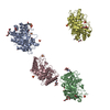
| ||||||||
|---|---|---|---|---|---|---|---|---|---|
| 1 | 
| ||||||||
| 2 | 
| ||||||||
| 3 | 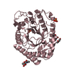
| ||||||||
| 4 | 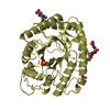
| ||||||||
| Unit cell |
|
-Components
-Protein / Sugars , 2 types, 8 molecules ABCD
| #1: Protein | Mass: 36841.055 Da / Num. of mol.: 4 Source method: isolated from a genetically manipulated source Details: 2-N-ACETYL-BETA-D-GLUCOSE(RESIDUE 601) LINKED TO ASN 111 IN THE FOUR MOLECULES Source: (gene. exp.) THIELAVIA HETEROTHALLICA (fungus)Description: MYCELIOPHTHORA THERMOPHILA IS THE ANAMORPH NAME WHILST THIELAVIA HETEROTHALLIC IS THE TELEOMORPH NAME Production host: References: UniProt: P83692*PLUS, arabinogalactan endo-beta-1,4-galactanase #2: Sugar | ChemComp-NAG /  Type: D-saccharide, beta linking / Mass: 221.208 Da / Num. of mol.: 4 Type: D-saccharide, beta linking / Mass: 221.208 Da / Num. of mol.: 4Source method: isolated from a genetically manipulated source Formula: C8H15NO6 |
|---|
-Non-polymers , 4 types, 668 molecules 


| #3: Chemical | ChemComp-SO4 /  Mass: 96.063 Da / Num. of mol.: 11 / Source method: obtained synthetically / Formula: SO4 Mass: 96.063 Da / Num. of mol.: 11 / Source method: obtained synthetically / Formula: SO4#4: Chemical | ChemComp-PEG /  Mass: 106.120 Da / Num. of mol.: 13 / Source method: obtained synthetically / Formula: C4H10O3 Mass: 106.120 Da / Num. of mol.: 13 / Source method: obtained synthetically / Formula: C4H10O3#5: Chemical | ChemComp-EPE / |  Mass: 238.305 Da / Num. of mol.: 1 / Source method: obtained synthetically / Formula: C8H18N2O4S / Comment: pH buffer*YM Mass: 238.305 Da / Num. of mol.: 1 / Source method: obtained synthetically / Formula: C8H18N2O4S / Comment: pH buffer*YM#6: Water | ChemComp-HOH / | Mass: 18.015 Da / Num. of mol.: 643 / Source method: isolated from a natural source / Formula: H2O |
|---|
-Details
| Has protein modification | Y |
|---|
-Experimental details
-Experiment
| Experiment | Method: X-RAY DIFFRACTION / Number of used crystals: 1 |
|---|
- Sample preparation
| Crystal | Density Matthews: 2.43 Å3/Da / Density % sol: 49.38 % | ||||||||||||||||||||||||||||||
|---|---|---|---|---|---|---|---|---|---|---|---|---|---|---|---|---|---|---|---|---|---|---|---|---|---|---|---|---|---|---|---|
| Crystal grow | pH: 7.5 Details: 30% PEG4000, 0.2 M AMMONIUM SULPHATE, 0.1M HEPES PH=7.5, pH 7.50 | ||||||||||||||||||||||||||||||
| Crystal grow | *PLUS pH: 7.5 / Method: vapor diffusion, hanging drop | ||||||||||||||||||||||||||||||
| Components of the solutions | *PLUS
|
-Data collection
| Diffraction | Mean temperature: 100 K |
|---|---|
| Diffraction source | Source: SYNCHROTRON / Site: MAX II  / Beamline: I711 / Wavelength: 1.07 / Beamline: I711 / Wavelength: 1.07 |
| Detector | Date: Nov 15, 2001 |
| Radiation | Protocol: SINGLE WAVELENGTH / Monochromatic (M) / Laue (L): M / Scattering type: x-ray |
| Radiation wavelength | Wavelength: 1.07 Å / Relative weight: 1 |
| Reflection | Resolution: 1.88→20 Å / Num. obs: 114360 / % possible obs: 97.6 % / Observed criterion σ(I): 2 / Redundancy: 4.4 % / Biso Wilson estimate: 12 Å2 / Rmerge(I) obs: 0.076 |
| Reflection shell | Resolution: 1.88→1.95 Å / Rmerge(I) obs: 0.417 / % possible all: 89 |
| Reflection | *PLUS Highest resolution: 1.88 Å / Lowest resolution: 20 Å / Rmerge(I) obs: 0.076 |
| Reflection shell | *PLUS % possible obs: 89 % / Rmerge(I) obs: 0.417 / Mean I/σ(I) obs: 61.3 |
- Processing
| Software |
| ||||||||||||||||||||||||||||||||||||||||||||||||||||||||||||
|---|---|---|---|---|---|---|---|---|---|---|---|---|---|---|---|---|---|---|---|---|---|---|---|---|---|---|---|---|---|---|---|---|---|---|---|---|---|---|---|---|---|---|---|---|---|---|---|---|---|---|---|---|---|---|---|---|---|---|---|---|---|
| Refinement | Method to determine structure: OTHER / Resolution: 1.87→19.84 Å / Rfactor Rfree error: 0.002 / Data cutoff high absF: 381740.82 / Isotropic thermal model: RESTRAINED / Cross valid method: THROUGHOUT / σ(F): 0
| ||||||||||||||||||||||||||||||||||||||||||||||||||||||||||||
| Solvent computation | Solvent model: FLAT MODEL / Bsol: 40.4333 Å2 / ksol: 0.393179 e/Å3 | ||||||||||||||||||||||||||||||||||||||||||||||||||||||||||||
| Displacement parameters | Biso mean: 16.9 Å2
| ||||||||||||||||||||||||||||||||||||||||||||||||||||||||||||
| Refine analyze |
| ||||||||||||||||||||||||||||||||||||||||||||||||||||||||||||
| Refinement step | Cycle: LAST / Resolution: 1.87→19.84 Å
| ||||||||||||||||||||||||||||||||||||||||||||||||||||||||||||
| Refine LS restraints |
| ||||||||||||||||||||||||||||||||||||||||||||||||||||||||||||
| LS refinement shell | Resolution: 1.87→1.99 Å / Rfactor Rfree error: 0.007 / Total num. of bins used: 6
| ||||||||||||||||||||||||||||||||||||||||||||||||||||||||||||
| Xplor file |
| ||||||||||||||||||||||||||||||||||||||||||||||||||||||||||||
| Refinement | *PLUS Highest resolution: 1.88 Å / Lowest resolution: 20 Å / σ(F): 0 / % reflection Rfree: 10 % / Rfactor obs: 0.194 / Rfactor Rfree: 0.208 / Rfactor Rwork: 0.194 | ||||||||||||||||||||||||||||||||||||||||||||||||||||||||||||
| Solvent computation | *PLUS | ||||||||||||||||||||||||||||||||||||||||||||||||||||||||||||
| Displacement parameters | *PLUS | ||||||||||||||||||||||||||||||||||||||||||||||||||||||||||||
| Refine LS restraints | *PLUS
| ||||||||||||||||||||||||||||||||||||||||||||||||||||||||||||
| LS refinement shell | *PLUS Rfactor Rfree: 0.257 / Rfactor Rwork: 0.243 |
