 Movie
Movie Controller

[English] 日本語
 Yorodumi
- PDB-1hfe: 1.6 A RESOLUTION STRUCTURE OF THE FE-ONLY HYDROGENASE FROM DESULF... -
+ Open data

- Basic information
| Entry | Database: PDB / ID: 1hfe | ||||||
|---|---|---|---|---|---|---|---|
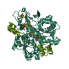
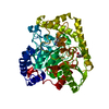
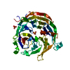
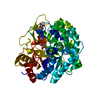
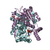
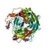
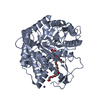
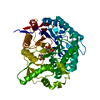
| Title | 1.6 A RESOLUTION STRUCTURE OF THE FE-ONLY HYDROGENASE FROM DESULFOVIBRIO DESULFURICANS | ||||||
 Components Components | (PROTEIN (FE-ONLY HYDROGENASE (E.C.1.18.99.1) ...) x 2 | ||||||
 Keywords Keywords | HYDROGENASE / FE-ONLY HYDROGENASE / HYDROGENE METABOLISM / PERIPLASM | ||||||
| Function / homology |  Function and homology information Function and homology informationferredoxin hydrogenase / ferredoxin hydrogenase activity / iron-sulfur cluster binding / 4 iron, 4 sulfur cluster binding / electron transfer activity / periplasmic space / iron ion binding Similarity search - Function | ||||||
| Biological species |  Desulfovibrio vulgaris subsp. vulgaris str. Hildenborough (bacteria) Desulfovibrio vulgaris subsp. vulgaris str. Hildenborough (bacteria) | ||||||
| Method |  X-RAY DIFFRACTION / SYNCHROTRON / MAD / Resolution: 1.6 Å X-RAY DIFFRACTION / SYNCHROTRON / MAD / Resolution: 1.6 Å | ||||||
 Authors Authors | Nicolet, Y. / Piras, C. / Legrand, P. / Hatchikian, E.C. / Fontecilla-Camps, J.C. | ||||||
 Citation Citation | Journal: Structure Fold.Des. / Year: 1999 Title: Desulfovibrio desulfuricans iron hydrogenase: the structure shows unusual coordination to an active site Fe binuclear center. Authors: Nicolet, Y. / Piras, C. / Legrand, P. / Hatchikian, C.E. / Fontecilla-Camps, J.C. | ||||||
| History |
|
- Structure visualization
| Structure viewer | Molecule: MolmilJmol/JSmol |
|---|
- Downloads & links
-Download
| PDBx/mmCIF format | 1hfe.cif.gz | 238.3 KB | Display | PDBx/mmCIF format |
|---|---|---|---|---|
| PDB format | pdb1hfe.ent.gz | 185.7 KB | Display | PDB format |
| PDBx/mmJSON format | 1hfe.json.gz | Tree view | PDBx/mmJSON format | |
| Others |  Other downloads Other downloads |
-Validation report
| Arichive directory | https://data.pdbj.org/pub/pdb/validation_reports/hf/1hfeftp://data.pdbj.org/pub/pdb/validation_reports/hf/1hfe | HTTPS FTP |
|---|
-Related structure data
| Similar structure data |
|---|


-Links
 PDBj
PDBj


- Assembly
| Deposited unit | 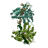
| ||||||||
|---|---|---|---|---|---|---|---|---|---|
| 1 | 
| ||||||||
| 2 | 
| ||||||||
| 3 |
| ||||||||
| Unit cell |
|
-Components
-PROTEIN (FE-ONLY HYDROGENASE (E.C.1.18.99.1) ... , 2 types, 4 molecules STLM
| #1: Protein | Mass: 13646.787 Da / Num. of mol.: 2 / Source method: isolated from a natural source Details: THE FE-ONLY HYDROGENASE FROM DESULFOVIBRIO DESULFURICANS HAS EXACTLY THE SAME SEQUENCE AS THE FE-ONLY HYDROGENASE FROM DESULFOVIBRIO VULGARIS (STRAIN HILDENBOROUGH) Source: (natural) Desulfovibrio vulgaris subsp. vulgaris str. Hildenborough (bacteria)Cellular location: PERIPLASM / Species: Desulfovibrio vulgaris / Strain: Hildenborough / ATCC 29579 / NCIMB 8303 / References: UniProt: P07603, 1.18.99.1 #2: Protein | Mass: 46011.133 Da / Num. of mol.: 2 / Source method: isolated from a natural source Details: THE FE-ONLY HYDROGENASE FROM DESULFOVIBRIO DESULFURICANS HAS EXACTLY THE SAME SEQUENCE AS THE FE-ONLY HYDROGENASE FROM DESULFOVIBRIO VULGARIS (STRAIN HILDENBOROUGH) Source: (natural) Desulfovibrio vulgaris subsp. vulgaris str. Hildenborough (bacteria)Cellular location: PERIPLASM / Species: Desulfovibrio vulgaris / Strain: Hildenborough / ATCC 29579 / NCIMB 8303 / References: UniProt: P07598, 1.18.99.1 |
|---|
-Non-polymers , 8 types, 1202 molecules 


| #3: Chemical | ChemComp-ZN /  Mass: 65.409 Da / Num. of mol.: 1 / Source method: obtained synthetically / Formula: Zn Mass: 65.409 Da / Num. of mol.: 1 / Source method: obtained synthetically / Formula: Zn | ||||||||||||
|---|---|---|---|---|---|---|---|---|---|---|---|---|---|
| #4: Chemical | ChemComp-FE2 /  Mass: 55.845 Da / Num. of mol.: 4 / Source method: obtained synthetically / Formula: Fe Mass: 55.845 Da / Num. of mol.: 4 / Source method: obtained synthetically / Formula: Fe#5: Chemical | ChemComp-CYN /  Mass: 26.017 Da / Num. of mol.: 4 / Source method: obtained synthetically / Formula: CN Mass: 26.017 Da / Num. of mol.: 4 / Source method: obtained synthetically / Formula: CN#6: Chemical | ChemComp-SF4 /  Mass: 351.640 Da / Num. of mol.: 6 / Source method: obtained synthetically / Formula: Fe4S4 Mass: 351.640 Da / Num. of mol.: 6 / Source method: obtained synthetically / Formula: Fe4S4#7: Chemical |  Mass: 108.226 Da / Num. of mol.: 2 / Source method: obtained synthetically / Formula: C3H8S2 Mass: 108.226 Da / Num. of mol.: 2 / Source method: obtained synthetically / Formula: C3H8S2#8: Chemical | ChemComp-CMO /  Mass: 28.010 Da / Num. of mol.: 4 / Source method: obtained synthetically / Formula: CO Mass: 28.010 Da / Num. of mol.: 4 / Source method: obtained synthetically / Formula: CO#9: Chemical |  Type: L-peptide linking / Mass: 121.158 Da / Num. of mol.: 2 / Source method: obtained synthetically / Formula: C3H7NO2S Type: L-peptide linking / Mass: 121.158 Da / Num. of mol.: 2 / Source method: obtained synthetically / Formula: C3H7NO2S#10: Water | ChemComp-HOH / | Mass: 18.015 Da / Num. of mol.: 1179 / Source method: isolated from a natural source / Formula: H2O |
-Experimental details
-Experiment
| Experiment | Method: X-RAY DIFFRACTION / Number of used crystals: 1 |
|---|
- Sample preparation
| Crystal | Density Matthews: 2.32 Å3/Da / Density % sol: 35 % | ||||||||||||||||||||||||
|---|---|---|---|---|---|---|---|---|---|---|---|---|---|---|---|---|---|---|---|---|---|---|---|---|---|
| Crystal grow | pH: 7.6 / Details: pH 7.60 | ||||||||||||||||||||||||
| Crystal grow | *PLUS Method: vapor diffusion, hanging drop / pH: 5 | ||||||||||||||||||||||||
| Components of the solutions | *PLUS
|
-Data collection
| Diffraction | Mean temperature: 100 K |
|---|---|
| Diffraction source | Source: SYNCHROTRON / Site: ESRF  / Beamline: ID14-3 / Wavelength: 0.947375 / Beamline: ID14-3 / Wavelength: 0.947375 |
| Detector | Type: MARRESEARCH / Detector: CCD / Date: May 1, 1998 / Details: MIRROR |
| Radiation | Monochromator: SI(111) / Protocol: SINGLE WAVELENGTH / Monochromatic (M) / Laue (L): M / Scattering type: x-ray |
| Radiation wavelength | Wavelength: 0.947375 Å / Relative weight: 1 |
| Reflection | Resolution: 1.51→21 Å / Num. obs: 158363 / % possible obs: 94.3 % / Redundancy: 6.4 % / Biso Wilson estimate: 14.27 Å2 / Rsym value: 0.071 / Net I/σ(I): 4.8 |
| Reflection shell | Resolution: 1.51→1.6 Å / Redundancy: 5.5 % / Mean I/σ(I) obs: 1.1 / Rsym value: 0.452 / % possible all: 89.2 |
| Reflection | *PLUS Rmerge(I) obs: 0.071 |
| Reflection shell | *PLUS % possible obs: 89.2 % / Rmerge(I) obs: 0.452 |
- Processing
| Software |
| ||||||||||||||||||||||||||||||||||||||||||||||||||||||||||||||||||||||||||||||||
|---|---|---|---|---|---|---|---|---|---|---|---|---|---|---|---|---|---|---|---|---|---|---|---|---|---|---|---|---|---|---|---|---|---|---|---|---|---|---|---|---|---|---|---|---|---|---|---|---|---|---|---|---|---|---|---|---|---|---|---|---|---|---|---|---|---|---|---|---|---|---|---|---|---|---|---|---|---|---|---|---|---|
| Refinement | Method to determine structure: MAD / Resolution: 1.6→7 Å / Rfactor Rfree error: 0.002 / Isotropic thermal model: RESTRAINED / Cross valid method: THROUGHOUT / σ(F): 2 Details: THE 2 MOLECULES OF THE ASYMMETRIC UNIT WERE NEVER CONSTRAINT WITH THE NON CRYSTALLOGRAPHIC SYMMETRY DURING REFINEMENT.
| ||||||||||||||||||||||||||||||||||||||||||||||||||||||||||||||||||||||||||||||||
| Displacement parameters | Biso mean: 16.52 Å2
| ||||||||||||||||||||||||||||||||||||||||||||||||||||||||||||||||||||||||||||||||
| Refinement step | Cycle: LAST / Resolution: 1.6→7 Å
| ||||||||||||||||||||||||||||||||||||||||||||||||||||||||||||||||||||||||||||||||
| Refine LS restraints |
| ||||||||||||||||||||||||||||||||||||||||||||||||||||||||||||||||||||||||||||||||
| LS refinement shell | Resolution: 1.6→1.67 Å / Rfactor Rfree error: 0.01 / Total num. of bins used: 8
| ||||||||||||||||||||||||||||||||||||||||||||||||||||||||||||||||||||||||||||||||
| Xplor file |
| ||||||||||||||||||||||||||||||||||||||||||||||||||||||||||||||||||||||||||||||||
| Software | *PLUS Name: X-PLOR / Version: 3.8 / Classification: refinement | ||||||||||||||||||||||||||||||||||||||||||||||||||||||||||||||||||||||||||||||||
| Refine LS restraints | *PLUS
|
