 Movie
Movie Controller


+ Open data

- Basic information
| Entry | Database: PDB / ID: 1gvg | ||||||
|---|---|---|---|---|---|---|---|
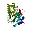
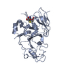
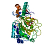
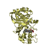
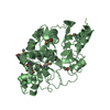
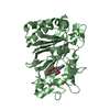
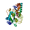
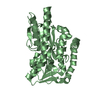
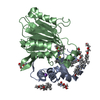
| Title | Crystal Structure of Clavaminate Synthase with Nitric Oxide | ||||||
 Components Components | CLAVAMINATE SYNTHASE 1 | ||||||
 Keywords Keywords | OXIDOREDUCTASE / LYASE / OXYGENASE / TRIFUNCTIONAL ENZYME / CLAVAMINATE SYNTHASE 1 / JELLY ROLL / NITRIC OXIDE | ||||||
| Function / homology |  Function and homology information Function and homology informationclavaminate synthase / clavaminate synthase activity / clavulanic acid biosynthetic process / antibiotic biosynthetic process / iron ion binding Similarity search - Function | ||||||
| Biological species |  STREPTOMYCES CLAVULIGERUS (bacteria) STREPTOMYCES CLAVULIGERUS (bacteria) | ||||||
| Method |  X-RAY DIFFRACTION / MOLECULAR REPLACEMENT / Resolution: 1.54 Å X-RAY DIFFRACTION / MOLECULAR REPLACEMENT / Resolution: 1.54 Å | ||||||
 Authors Authors | Zhang, Z.H. / Ren, J. / McKinnon, C.H. / Clifton, I.J. / Harlos, K. / Schofield, C.J. | ||||||
 Citation Citation | Journal: FEBS Lett. / Year: 2002 Title: Crystal Structure of a Clavaminate Synthase-Fe(II) -2-Oxoglutarate-Substrate-No Complex: Evidence for Metal Centered Rearrangements Authors: Zhang, Z.H. / Ren, J. / Harlos, K. / McKinnon, C.H. / Clifton, I.J. / Schofield, C.J. #1: Journal: Nat.Struct.Biol. / Year: 2000Title: Structural Origins of the Selectivity of the Trifunctional Oxygenase Clavaminic Acid Synthase Authors: Zhang, Z.H. / Ren, J. / Stammers, D.K. / Baldwin, J.E. / Harlos, K. / Schofield, C.J. | ||||||
| History |
| ||||||
| Remark 700 | SHEET THE SHEET STRUCTURE OF THIS MOLECULE IS BIFURCATED. IN ORDER TO REPRESENT THIS FEATURE IN ... SHEET THE SHEET STRUCTURE OF THIS MOLECULE IS BIFURCATED. IN ORDER TO REPRESENT THIS FEATURE IN THE SHEET RECORDS BELOW, TWO SHEETS ARE DEFINED. |
- Structure visualization
| Structure viewer | Molecule: MolmilJmol/JSmol |
|---|
- Downloads & links
-Download
| PDBx/mmCIF format | 1gvg.cif.gz | 86.4 KB | Display | PDBx/mmCIF format |
|---|---|---|---|---|
| PDB format | pdb1gvg.ent.gz | 62.6 KB | Display | PDB format |
| PDBx/mmJSON format | 1gvg.json.gz | Tree view | PDBx/mmJSON format | |
| Others |  Other downloads Other downloads |
-Validation report
| Arichive directory | https://data.pdbj.org/pub/pdb/validation_reports/gv/1gvgftp://data.pdbj.org/pub/pdb/validation_reports/gv/1gvg | HTTPS FTP |
|---|
-Related structure data
| Related structure data | 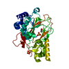 1ds0S S: Starting model for refinement |
|---|---|
| Similar structure data |


-Links
 PDBj
PDBj

- Assembly
| Deposited unit | 
| ||||||||
|---|---|---|---|---|---|---|---|---|---|
| 1 |
| ||||||||
| Unit cell |
|
-Components
-Protein , 1 types, 1 molecules A
| #1: Protein | Mass: 35415.785 Da / Num. of mol.: 1 Source method: isolated from a genetically manipulated source Source: (gene. exp.) STREPTOMYCES CLAVULIGERUS (bacteria) / Plasmid: PET11A / Production host: |
|---|
-Non-polymers , 6 types, 399 molecules 


| #2: Chemical | ChemComp-FE /  Mass: 55.845 Da / Num. of mol.: 1 / Source method: obtained synthetically / Formula: Fe Mass: 55.845 Da / Num. of mol.: 1 / Source method: obtained synthetically / Formula: Fe | ||||||
|---|---|---|---|---|---|---|---|
| #3: Chemical | ChemComp-AKG /  Mass: 146.098 Da / Num. of mol.: 1 / Source method: obtained synthetically / Formula: C5H6O5 Mass: 146.098 Da / Num. of mol.: 1 / Source method: obtained synthetically / Formula: C5H6O5 | ||||||
| #4: Chemical |  Mass: 96.063 Da / Num. of mol.: 3 / Source method: obtained synthetically / Formula: SO4 Mass: 96.063 Da / Num. of mol.: 3 / Source method: obtained synthetically / Formula: SO4#5: Chemical | ChemComp-PCX / |  Mass: 228.248 Da / Num. of mol.: 1 / Source method: obtained synthetically / Formula: C9H16N4O3 Mass: 228.248 Da / Num. of mol.: 1 / Source method: obtained synthetically / Formula: C9H16N4O3#6: Chemical | ChemComp-HOA / |  Mass: 33.030 Da / Num. of mol.: 1 / Source method: obtained synthetically / Formula: H3NO Mass: 33.030 Da / Num. of mol.: 1 / Source method: obtained synthetically / Formula: H3NO#7: Water | ChemComp-HOH / | Mass: 18.015 Da / Num. of mol.: 392 / Source method: isolated from a natural source / Formula: H2O |
-Experimental details
-Experiment
| Experiment | Method: X-RAY DIFFRACTION / Number of used crystals: 1 |
|---|
- Sample preparation
| Crystal | Density Matthews: 2.2 Å3/Da / Density % sol: 43.65 % | ||||||||||||||||||||||||||||||
|---|---|---|---|---|---|---|---|---|---|---|---|---|---|---|---|---|---|---|---|---|---|---|---|---|---|---|---|---|---|---|---|
| Crystal grow | pH: 7.6 / Details: pH 7.60 | ||||||||||||||||||||||||||||||
| Crystal grow | *PLUS Temperature: 20 ℃ / pH: 7.5 / Method: vapor diffusion, hanging drop | ||||||||||||||||||||||||||||||
| Components of the solutions | *PLUS
|
-Data collection
| Diffraction | Mean temperature: 100 K |
|---|---|
| Diffraction source | Source: ROTATING ANODE / Type: RIGAKU RU200H / Wavelength: 1.5418 |
| Detector | Type: MAR scanner 345 mm plate / Detector: IMAGE PLATE / Date: Feb 15, 2001 |
| Radiation | Protocol: SINGLE WAVELENGTH / Monochromatic (M) / Laue (L): M / Scattering type: x-ray |
| Radiation wavelength | Wavelength: 1.5418 Å / Relative weight: 1 |
| Reflection | Resolution: 1.54→30 Å / Num. obs: 46381 / % possible obs: 97.7 % / Redundancy: 13.2 % / Biso Wilson estimate: 19.8 Å2 / Rmerge(I) obs: 0.054 / Net I/σ(I): 29.1 |
| Reflection shell | Resolution: 1.54→1.6 Å / Redundancy: 4.46 % / Rmerge(I) obs: 0.298 / Mean I/σ(I) obs: 3.8 / % possible all: 86 |
| Reflection | *PLUS Lowest resolution: 30 Å / Num. measured all: 301822 |
| Reflection shell | *PLUS % possible obs: 86 % / Num. unique obs: 4038 |
- Processing
| Software |
| ||||||||||||||||||||||||||||||||||||||||||||||||||||||||||||
|---|---|---|---|---|---|---|---|---|---|---|---|---|---|---|---|---|---|---|---|---|---|---|---|---|---|---|---|---|---|---|---|---|---|---|---|---|---|---|---|---|---|---|---|---|---|---|---|---|---|---|---|---|---|---|---|---|---|---|---|---|---|
| Refinement | Method to determine structure: MOLECULAR REPLACEMENT Starting model: PDB ENTRY 1DS0 Resolution: 1.54→30 Å / Cross valid method: THROUGHOUT / σ(F): 0
| ||||||||||||||||||||||||||||||||||||||||||||||||||||||||||||
| Solvent computation | Bsol: 46.29 Å2 / ksol: 0.4 e/Å3 | ||||||||||||||||||||||||||||||||||||||||||||||||||||||||||||
| Displacement parameters |
| ||||||||||||||||||||||||||||||||||||||||||||||||||||||||||||
| Refinement step | Cycle: LAST / Resolution: 1.54→30 Å
| ||||||||||||||||||||||||||||||||||||||||||||||||||||||||||||
| Refine LS restraints |
| ||||||||||||||||||||||||||||||||||||||||||||||||||||||||||||
| Xplor file |
| ||||||||||||||||||||||||||||||||||||||||||||||||||||||||||||
| Refinement | *PLUS Lowest resolution: 30 Å / % reflection Rfree: 5 % / Rfactor Rfree: 0.2074 / Rfactor Rwork: 0.1813 | ||||||||||||||||||||||||||||||||||||||||||||||||||||||||||||
| Solvent computation | *PLUS | ||||||||||||||||||||||||||||||||||||||||||||||||||||||||||||
| Displacement parameters | *PLUS | ||||||||||||||||||||||||||||||||||||||||||||||||||||||||||||
| Refine LS restraints | *PLUS
|
