 Movie
Movie Controller


+ Open data

- Basic information
| Entry | Database: PDB / ID: 6yrd | ||||||
|---|---|---|---|---|---|---|---|
| Title | SFX structure of dye-type peroxidase DtpB in the ferryl state | ||||||
 Components Components | Putative iron-dependent peroxidase | ||||||
 Keywords Keywords | OXIDOREDUCTASE / peroxidase / dye-decolourising / serial femtosecond crystallography / damage-free / ferryl | ||||||
| Function / homology |  Function and homology information Function and homology information | ||||||
| Biological species |  Streptomyces lividans 1326 (bacteria) Streptomyces lividans 1326 (bacteria) | ||||||
| Method |  X-RAY DIFFRACTION / FREE ELECTRON LASER / MOLECULAR REPLACEMENT / Resolution: 1.75 Å X-RAY DIFFRACTION / FREE ELECTRON LASER / MOLECULAR REPLACEMENT / Resolution: 1.75 Å | ||||||
 Authors Authors | Lucic, M. / Axford, D.A. / Owen, R.L. / Worrall, J.A.R. / Hough, M.A. | ||||||
| Funding support |  United Kingdom, 1items United Kingdom, 1items
| ||||||
 Citation Citation | Journal: Angew.Chem.Int.Ed.Engl. / Year: 2020 Title: Serial Femtosecond Zero Dose Crystallography Captures a Water-Free Distal Heme Site in a Dye-Decolorising Peroxidase to Reveal a Catalytic Role for an Arginine in Fe IV =O Formation. Authors: Lucic, M. / Svistunenko, D.A. / Wilson, M.T. / Chaplin, A.K. / Davy, B. / Ebrahim, A. / Axford, D. / Tosha, T. / Sugimoto, H. / Owada, S. / Dworkowski, F.S.N. / Tews, I. / Owen, R.L. / ...Authors: Lucic, M. / Svistunenko, D.A. / Wilson, M.T. / Chaplin, A.K. / Davy, B. / Ebrahim, A. / Axford, D. / Tosha, T. / Sugimoto, H. / Owada, S. / Dworkowski, F.S.N. / Tews, I. / Owen, R.L. / Hough, M.A. / Worrall, J.A.R. | ||||||
| History |
|
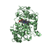
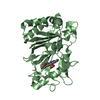
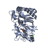
- Structure visualization
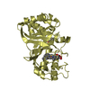
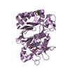
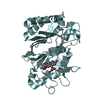
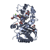
| Structure viewer | Molecule: MolmilJmol/JSmol |
|---|
- Downloads & links
-Download
| PDBx/mmCIF format | 6yrd.cif.gz | 383.7 KB | Display | PDBx/mmCIF format |
|---|---|---|---|---|
| PDB format | pdb6yrd.ent.gz | 311.7 KB | Display | PDB format |
| PDBx/mmJSON format | 6yrd.json.gz | Tree view | PDBx/mmJSON format | |
| Others |  Other downloads Other downloads |
-Validation report
| Arichive directory | https://data.pdbj.org/pub/pdb/validation_reports/yr/6yrdftp://data.pdbj.org/pub/pdb/validation_reports/yr/6yrd | HTTPS FTP |
|---|
-Related structure data


-Links
 PDBj
PDBj

- Assembly
| Deposited unit | 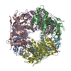
| ||||||||
|---|---|---|---|---|---|---|---|---|---|
| 1 | 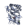
| ||||||||
| 2 | 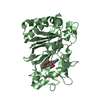
| ||||||||
| 3 | 
| ||||||||
| 4 | 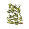
| ||||||||
| 5 | 
| ||||||||
| 6 | 
| ||||||||
| Unit cell |
|
-Components
-Protein , 1 types, 6 molecules ABCDEF
| #1: Protein | Mass: 34172.223 Da / Num. of mol.: 6 Source method: isolated from a genetically manipulated source Source: (gene. exp.) Streptomyces lividans 1326 (bacteria) / Gene: SAMN05428941_7146 / Plasmid: pET28a / Production host: |
|---|
-Non-polymers , 5 types, 895 molecules 


| #2: Chemical | ChemComp-HEM /  Mass: 616.487 Da / Num. of mol.: 6 / Source method: obtained synthetically / Formula: C34H32FeN4O4 Mass: 616.487 Da / Num. of mol.: 6 / Source method: obtained synthetically / Formula: C34H32FeN4O4#3: Chemical |  Mass: 24.305 Da / Num. of mol.: 2 / Source method: obtained synthetically / Formula: Mg Mass: 24.305 Da / Num. of mol.: 2 / Source method: obtained synthetically / Formula: Mg#4: Chemical | ChemComp-O /  Mass: 15.999 Da / Num. of mol.: 6 / Source method: obtained synthetically / Formula: O / Feature type: SUBJECT OF INVESTIGATION Mass: 15.999 Da / Num. of mol.: 6 / Source method: obtained synthetically / Formula: O / Feature type: SUBJECT OF INVESTIGATION#5: Chemical | ChemComp-PG4 / |  Mass: 194.226 Da / Num. of mol.: 1 / Source method: obtained synthetically / Formula: C8H18O5 / Comment: precipitant*YM Mass: 194.226 Da / Num. of mol.: 1 / Source method: obtained synthetically / Formula: C8H18O5 / Comment: precipitant*YM#6: Water | ChemComp-HOH / | Mass: 18.015 Da / Num. of mol.: 880 / Source method: isolated from a natural source / Formula: H2O |
|---|
-Details
| Has ligand of interest | Y |
|---|---|
| Has protein modification | N |
-Experimental details
-Experiment
| Experiment | Method: X-RAY DIFFRACTION / Number of used crystals: 1 |
|---|
- Sample preparation
| Crystal | Density Matthews: 2.51 Å3/Da / Density % sol: 50.92 % / Description: Microcrystals |
|---|---|
| Crystal grow | Temperature: 293 K / Method: batch mode / pH: 7.5 Details: 6-10 mg/mL of protein in 50mM Sodium acetate, 150mM NaCl pH 5 was mixed with 150 mM MgCl2, 150 mM HEPES, 20% PEG 4000 pH 7.5 |
-Data collection
| Diffraction | Mean temperature: 301 K / Serial crystal experiment: Y |
|---|---|
| Diffraction source | Source: FREE ELECTRON LASER / Site: SACLA  / Beamline: BL2 / Wavelength: 1.13 Å / Beamline: BL2 / Wavelength: 1.13 Å |
| Detector | Type: MPCCD / Detector: CCD / Date: Jun 26, 2019 |
| Radiation | Protocol: SINGLE WAVELENGTH / Monochromatic (M) / Laue (L): M / Scattering type: x-ray |
| Radiation wavelength | Wavelength: 1.13 Å / Relative weight: 1 |
| Reflection | Resolution: 1.75→102 Å / Num. obs: 207440 / % possible obs: 100 % / Redundancy: 1943 % / Biso Wilson estimate: 28.4 Å2 / CC1/2: 0.984 / R split: 0.109 / Net I/σ(I): 6.1 |
| Reflection shell | Resolution: 1.75→1.8 Å / Num. unique obs: 10274 / CC1/2: 0.423 / R split: 0.887 |
| Serial crystallography measurement | Pulse duration: 10 fsec. / Pulse energy: 156 µJ / Pulse photon energy: 11 keV |
| Serial crystallography sample delivery | Description: silicon chip / Method: fixed target |
| Serial crystallography sample delivery fixed target | Description: silicon chip / Details: 25600 apertures per chip |
- Processing
| Software |
| ||||||||||||||||||||||||||||||||||||||||||||||||||||||||||||
|---|---|---|---|---|---|---|---|---|---|---|---|---|---|---|---|---|---|---|---|---|---|---|---|---|---|---|---|---|---|---|---|---|---|---|---|---|---|---|---|---|---|---|---|---|---|---|---|---|---|---|---|---|---|---|---|---|---|---|---|---|---|
| Refinement | Method to determine structure: MOLECULAR REPLACEMENT Starting model: unpublished structure Resolution: 1.75→13 Å / Cor.coef. Fo:Fc: 0.971 / Cor.coef. Fo:Fc free: 0.959 / SU B: 2.594 / SU ML: 0.078 / SU R Cruickshank DPI: 0.1004 / Cross valid method: THROUGHOUT / σ(F): 0 / ESU R: 0.1 / ESU R Free: 0.099 Details: HYDROGENS HAVE BEEN ADDED IN THE RIDING POSITIONS U VALUES : REFINED INDIVIDUALLY
| ||||||||||||||||||||||||||||||||||||||||||||||||||||||||||||
| Solvent computation | Ion probe radii: 0.8 Å / Shrinkage radii: 0.8 Å / VDW probe radii: 1.2 Å | ||||||||||||||||||||||||||||||||||||||||||||||||||||||||||||
| Displacement parameters | Biso max: 105.34 Å2 / Biso mean: 33.628 Å2 / Biso min: 19.27 Å2
| ||||||||||||||||||||||||||||||||||||||||||||||||||||||||||||
| Refinement step | Cycle: final / Resolution: 1.75→13 Å
| ||||||||||||||||||||||||||||||||||||||||||||||||||||||||||||
| Refine LS restraints |
| ||||||||||||||||||||||||||||||||||||||||||||||||||||||||||||
| LS refinement shell | Resolution: 1.75→1.795 Å / Rfactor Rfree error: 0 / Total num. of bins used: 20
|
