 Movie
Movie Controller

[English] 日本語
 Yorodumi
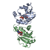
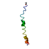
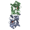
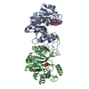
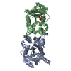
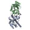
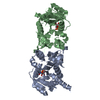
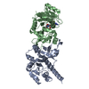
- PDB-1gqc: THE STRUCTURE OF CMP:2-KETO-3-DEOXY-MANNO-OCTONIC ACID SYNTHETASE... -
+ Open data

- Basic information
| Entry | Database: PDB / ID: 1gqc | ||||||
|---|---|---|---|---|---|---|---|
| Title | THE STRUCTURE OF CMP:2-KETO-3-DEOXY-MANNO-OCTONIC ACID SYNTHETASE COMPLEXED WITH CMP-Kdo at 100K | ||||||
 Components Components | 3-DEOXY-MANNO-OCTULOSONATE CYTIDYLYLTRANSFERASE | ||||||
 Keywords Keywords | TRANSFERASE / NUCLEOTIDYLTRANSFERASE / CMP-KDO SYNTHETASE / NUCLEOSIDE MONOPHOSPHATE GLYCOSIDES / LIPOPOLYSACCHARIDE BIOSYNTHESIS / SUGAR-ACTIVATING ENZYMES | ||||||
| Function / homology |  Function and homology information Function and homology information3-deoxy-manno-octulosonate cytidylyltransferase / 3-deoxy-manno-octulosonate cytidylyltransferase activity / CMP-keto-3-deoxy-D-manno-octulosonic acid biosynthetic process / lipopolysaccharide biosynthetic process / cytosol Similarity search - Function | ||||||
| Biological species |  | ||||||
| Method |  X-RAY DIFFRACTION / OTHER / Resolution: 2.6 Å X-RAY DIFFRACTION / OTHER / Resolution: 2.6 Å | ||||||
 Authors Authors | Jelakovic, S. / Schulz, G.E. | ||||||
 Citation Citation | Journal: Biochemistry / Year: 2002 Title: Catalytic Mechanism of Cmp:2-Keto-3-Deoxy-Manno-Octonic Acid Synthetase as Derived from Complexes with Reaction Educt and Product. Authors: Jelakovic, S. / Schulz, G.E. #1: Journal: J.Mol.Biol. / Year: 2001Title: The Structure of Cmp:2-Keto-3-Deoxy-Manno-Octonic Acid Synthetase and of its Complexes with Substrates and Substrate Analogs Authors: Jelakovic, S. / Schulz, G.E. #2: Journal: FEBS Lett. / Year: 1996Title: The Three-Dimensional Structure of Capsule-Specific Cmp:2-Keto-3-Deoxy-Manno-Octonic Acid Synthetase from Escherichia Coli Authors: Jelakovic, S. / Jann, K. / Schulz, G.E. | ||||||
| History |
| ||||||
| Remark 700 | SHEET THE SHEET STRUCTURE OF THIS MOLECULE IS BIFURCATED. IN ORDER TO REPRESENT THIS FEATURE IN ... SHEET THE SHEET STRUCTURE OF THIS MOLECULE IS BIFURCATED. IN ORDER TO REPRESENT THIS FEATURE IN THE SHEET RECORDS BELOW, TWO SHEETS ARE DEFINED. |
- Structure visualization
| Structure viewer | Molecule: MolmilJmol/JSmol |
|---|
- Downloads & links
-Download
| PDBx/mmCIF format | 1gqc.cif.gz | 112.3 KB | Display | PDBx/mmCIF format |
|---|---|---|---|---|
| PDB format | pdb1gqc.ent.gz | 86.3 KB | Display | PDB format |
| PDBx/mmJSON format | 1gqc.json.gz | Tree view | PDBx/mmJSON format | |
| Others |  Other downloads Other downloads |
-Validation report
| Arichive directory | https://data.pdbj.org/pub/pdb/validation_reports/gq/1gqcftp://data.pdbj.org/pub/pdb/validation_reports/gq/1gqc | HTTPS FTP |
|---|
-Related structure data


-Links
 PDBj
PDBj

- Assembly
| Deposited unit | 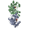
| ||||||||
|---|---|---|---|---|---|---|---|---|---|
| 1 |
| ||||||||
| Unit cell |
| ||||||||
| Noncrystallographic symmetry (NCS) | NCS oper: (Code: given Matrix: (-0.081444, 0.885712, -0.457036), Vector: |
-Components
| #1: Protein | Mass: 27059.912 Da / Num. of mol.: 2 Source method: isolated from a genetically manipulated source Source: (gene. exp.) References: UniProt: P42216, 3-deoxy-manno-octulosonate cytidylyltransferase #2: Chemical |   Mass: 24.305 Da / Num. of mol.: 2 / Source method: obtained synthetically / Formula: Mg Mass: 24.305 Da / Num. of mol.: 2 / Source method: obtained synthetically / Formula: Mg#3: Chemical | ChemComp-CMK / | 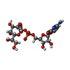  Mass: 543.373 Da / Num. of mol.: 1 / Source method: obtained synthetically / Formula: C17H26N3O15P Mass: 543.373 Da / Num. of mol.: 1 / Source method: obtained synthetically / Formula: C17H26N3O15P#4: Chemical | ChemComp-C5P / |   Mass: 323.197 Da / Num. of mol.: 1 / Source method: obtained synthetically / Formula: C9H14N3O8P Mass: 323.197 Da / Num. of mol.: 1 / Source method: obtained synthetically / Formula: C9H14N3O8P#5: Water | ChemComp-HOH / |  Mass: 18.015 Da / Num. of mol.: 267 / Source method: isolated from a natural source / Formula: H2O Mass: 18.015 Da / Num. of mol.: 267 / Source method: isolated from a natural source / Formula: H2O |
|---|
-Experimental details
-Experiment
| Experiment | Method: X-RAY DIFFRACTION |
|---|
- Sample preparation
| Crystal | Density Matthews: 2.62 Å3/Da / Density % sol: 52.97 % | ||||||||||||||||||||||||||||||||||||||||||||||||
|---|---|---|---|---|---|---|---|---|---|---|---|---|---|---|---|---|---|---|---|---|---|---|---|---|---|---|---|---|---|---|---|---|---|---|---|---|---|---|---|---|---|---|---|---|---|---|---|---|---|
| Crystal grow | pH: 9.4 / Details: pH 9.40 | ||||||||||||||||||||||||||||||||||||||||||||||||
| Crystal grow | *PLUS Temperature: 20 ℃ / Method: vapor diffusion, hanging drop / Details: Jelakovic, S., (2001) J.Mol.Biol., 312, 143. | ||||||||||||||||||||||||||||||||||||||||||||||||
| Components of the solutions | *PLUS
|
-Data collection
| Diffraction | Mean temperature: 100 K |
|---|---|
| Diffraction source | Source: ROTATING ANODE / Wavelength: 1.5418 |
| Radiation | Protocol: SINGLE WAVELENGTH / Monochromatic (M) / Laue (L): M / Scattering type: x-ray |
| Radiation wavelength | Wavelength: 1.5418 Å / Relative weight: 1 |
| Reflection | Resolution: 2.6→33 Å / Num. obs: 15622 / % possible obs: 88 % / Redundancy: 5.9 % / Rmerge(I) obs: 0.115 / Net I/σ(I): 6 |
| Reflection | *PLUS Lowest resolution: 33 Å / % possible obs: 88 % |
| Reflection shell | *PLUS Highest resolution: 2.57 Å / Lowest resolution: 2.7 Å / % possible obs: 81 % / Redundancy: 5.7 % / Num. unique obs: 2075 / Rmerge(I) obs: 0.33 / Mean I/σ(I) obs: 2.3 |
- Processing
| Software | Name: CNS / Classification: refinement | ||||||||||||||||||||||||||||||||||||||||||||||||||||||||||||
|---|---|---|---|---|---|---|---|---|---|---|---|---|---|---|---|---|---|---|---|---|---|---|---|---|---|---|---|---|---|---|---|---|---|---|---|---|---|---|---|---|---|---|---|---|---|---|---|---|---|---|---|---|---|---|---|---|---|---|---|---|---|
| Refinement | Method to determine structure: OTHER / Resolution: 2.6→24.44 Å / Cross valid method: THROUGHOUT / σ(F): 0
| ||||||||||||||||||||||||||||||||||||||||||||||||||||||||||||
| Refinement step | Cycle: LAST / Resolution: 2.6→24.44 Å
| ||||||||||||||||||||||||||||||||||||||||||||||||||||||||||||
| Refine LS restraints |
| ||||||||||||||||||||||||||||||||||||||||||||||||||||||||||||
| Refinement | *PLUS Lowest resolution: 25 Å / Rfactor Rfree: 0.24 | ||||||||||||||||||||||||||||||||||||||||||||||||||||||||||||
| Solvent computation | *PLUS | ||||||||||||||||||||||||||||||||||||||||||||||||||||||||||||
| Displacement parameters | *PLUS |
