 Movie
Movie Controller

[English] 日本語
 Yorodumi
- PDB-1gnl: Hybrid Cluster Protein from Desulfovibrio desulfuricans X-ray str... -
+ Open data

- Basic information
| Entry | Database: PDB / ID: 1gnl | |||||||||
|---|---|---|---|---|---|---|---|---|---|---|
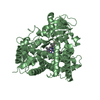
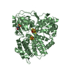
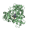
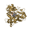
| Title | Hybrid Cluster Protein from Desulfovibrio desulfuricans X-ray structure at 1.25A resolution using synchrotron radiation at a wavelength of 0.933A | |||||||||
 Components Components | HYBRID CLUSTER PROTEIN | |||||||||
 Keywords Keywords | HYBRID CLUSTER PROTEIN / ANAEROBIC DESULFOVIBRIO DESULFURICANS / IRON ANOMALOUS | |||||||||
| Function / homology |  Function and homology information Function and homology information hydroxylamine reductase / hydroxylamine reductase activity / 4 iron, 4 sulfur cluster binding / metal ion binding / cytoplasm hydroxylamine reductase / hydroxylamine reductase activity / 4 iron, 4 sulfur cluster binding / metal ion binding / cytoplasmSimilarity search - Function | |||||||||
| Biological species |  DESULFOVIBRIO DESULFURICANS (bacteria) DESULFOVIBRIO DESULFURICANS (bacteria) | |||||||||
| Method | X-RAY DIFFRACTION / SYNCHROTRON / MOLECULAR REPLACEMENT / Resolution: 1.25 Å | |||||||||
 Authors Authors | Macedo, S. / Mitchell, E.P. / Romao, C.V. / Cooper, S.J. / Coelho, R. / Liu, M.Y. / Xavier, A.V. / Legall, J. / Bailey, S. / Garner, D.C. ...Macedo, S. / Mitchell, E.P. / Romao, C.V. / Cooper, S.J. / Coelho, R. / Liu, M.Y. / Xavier, A.V. / Legall, J. / Bailey, S. / Garner, D.C. / Hagen, W.R. / Teixeira, M. / Carrondo, M.A. / Lindley, P. | |||||||||
 Citation Citation | Journal: J.Biol.Inorg.Chem. / Year: 2002 Title: Hybrid Cluster Proteins (Hcps) from Desulfovibrio Desulfuricans Atcc 27774 and Desulfovibrio Vulgaris (Hildenborough): X-Ray Structures at 1.25 A Resolution Using Synchrotron Radiation. Authors: Macedo, S. / Mitchell, E.P. / Romao, C.V. / Cooper, S.J. / Coelho, R. / Liu, M.Y. / Xavier, A.V. / Legall, J. / Bailey, S. / Garner, D.C. / Hagen, W.R. / Teixeira, M. / Carrondo, M.A. / Lindley, P. | |||||||||
| History |
|
- Structure visualization
| Structure viewer | Molecule: MolmilJmol/JSmol |
|---|
- Downloads & links
-Download
| PDBx/mmCIF format | 1gnl.cif.gz | 250.2 KB | Display | PDBx/mmCIF format |
|---|---|---|---|---|
| PDB format | pdb1gnl.ent.gz | 199.2 KB | Display | PDB format |
| PDBx/mmJSON format | 1gnl.json.gz | Tree view | PDBx/mmJSON format | |
| Others |  Other downloads Other downloads |
-Validation report
| Arichive directory | https://data.pdbj.org/pub/pdb/validation_reports/gn/1gnlftp://data.pdbj.org/pub/pdb/validation_reports/gn/1gnl | HTTPS FTP |
|---|
-Related structure data
| Related structure data |  1gn9SC  1gntC S: Starting model for refinement C: citing same article ( |
|---|---|
| Similar structure data |

-Links
 PDBj
PDBj- Assembly
| Deposited unit | 
| ||||||||
|---|---|---|---|---|---|---|---|---|---|
| 1 | 
| ||||||||
| 2 | 
| ||||||||
| Unit cell |
|
-Components
| #1: Protein | Mass: 58595.879 Da / Num. of mol.: 2 / Source method: isolated from a natural source Details: CUBANE CLUSTER [4FE-4S] HYBRID CLUSTER [4FE-3S-3O] PERSULPHIDE BOND BETWEEN S7 (HYBRID CLUSTER) AND S OF CYS399 Source: (natural) DESULFOVIBRIO DESULFURICANS (bacteria) / References: UniProt: Q01770#2: Chemical | Iron–sulfur cluster  Mass: 351.640 Da / Num. of mol.: 2 / Source method: obtained synthetically / Formula: Fe4S4 Mass: 351.640 Da / Num. of mol.: 2 / Source method: obtained synthetically / Formula: Fe4S4#3: Chemical |   Mass: 367.573 Da / Num. of mol.: 2 / Source method: obtained synthetically / Formula: Fe4O3S3 Mass: 367.573 Da / Num. of mol.: 2 / Source method: obtained synthetically / Formula: Fe4O3S3#4: Chemical | ChemComp-ACT / | Acetate  Mass: 59.044 Da / Num. of mol.: 1 / Source method: obtained synthetically / Formula: C2H3O2 Mass: 59.044 Da / Num. of mol.: 1 / Source method: obtained synthetically / Formula: C2H3O2#5: Water | ChemComp-HOH / | Water Mass: 18.015 Da / Num. of mol.: 1562 / Source method: isolated from a natural source / Formula: H2O Mass: 18.015 Da / Num. of mol.: 1562 / Source method: isolated from a natural source / Formula: H2O |
|---|
-Experimental details
-Experiment
| Experiment | Method: X-RAY DIFFRACTION / Number of used crystals: 1 |
|---|
- Sample preparation
| Crystal | Density Matthews: 2.03 Å3/Da / Density % sol: 39 % | ||||||||||||||||||||||||||||||||||||||||||
|---|---|---|---|---|---|---|---|---|---|---|---|---|---|---|---|---|---|---|---|---|---|---|---|---|---|---|---|---|---|---|---|---|---|---|---|---|---|---|---|---|---|---|---|
| Crystal grow | Temperature: 277 K / pH: 6 / Details: 25% PEG 4000, 0.1M MES PH6.0, T=277K, pH 6.00 | ||||||||||||||||||||||||||||||||||||||||||
| Crystal grow | *PLUS Temperature: 293 K / pH: 7.6 / Method: vapor diffusion, sitting drop | ||||||||||||||||||||||||||||||||||||||||||
| Components of the solutions | *PLUS
|
-Data collection
| Diffraction | Mean temperature: 100 K |
|---|---|
| Diffraction source | Source: SYNCHROTRON / Site: ESRF  / Beamline: ID14-2 / Wavelength: 0.933 / Beamline: ID14-2 / Wavelength: 0.933 |
| Detector | Type: ADSC CCD / Detector: CCD / Date: May 15, 2001 / Details: TOROIDAL MIRROR |
| Radiation | Monochromator: DIAMOND (111) CRYSTAL / Protocol: SINGLE WAVELENGTH / Monochromatic (M) / Laue (L): M / Scattering type: x-ray |
| Radiation wavelength | Wavelength: 0.933 Å / Relative weight: 1 |
| Reflection | Resolution: 1.25→28.63 Å / Num. obs: 247243 / % possible obs: 94.9 % / Redundancy: 3.7 % / Rsym value: 0.054 / Net I/σ(I): 8 |
| Reflection shell | Resolution: 1.25→1.29 Å / Redundancy: 3.7 % / Mean I/σ(I) obs: 3.1 / Rsym value: 0.215 / % possible all: 85.3 |
| Reflection | *PLUS Num. measured all: 915955 / Rmerge(I) obs: 0.054 |
| Reflection shell | *PLUS % possible obs: 85.3 % / Rmerge(I) obs: 0.215 |
- Processing
| Software |
| ||||||||||||||||||||||||||||||||||||||||||||||||||||||||||||||||||||||||||||||||||||||||||||||||||||||||||||||||||||||||||||||||||||||||||||||||||||||||||||||||||||||||||||||||||||||
|---|---|---|---|---|---|---|---|---|---|---|---|---|---|---|---|---|---|---|---|---|---|---|---|---|---|---|---|---|---|---|---|---|---|---|---|---|---|---|---|---|---|---|---|---|---|---|---|---|---|---|---|---|---|---|---|---|---|---|---|---|---|---|---|---|---|---|---|---|---|---|---|---|---|---|---|---|---|---|---|---|---|---|---|---|---|---|---|---|---|---|---|---|---|---|---|---|---|---|---|---|---|---|---|---|---|---|---|---|---|---|---|---|---|---|---|---|---|---|---|---|---|---|---|---|---|---|---|---|---|---|---|---|---|---|---|---|---|---|---|---|---|---|---|---|---|---|---|---|---|---|---|---|---|---|---|---|---|---|---|---|---|---|---|---|---|---|---|---|---|---|---|---|---|---|---|---|---|---|---|---|---|---|---|
| Refinement | Method to determine structure: MOLECULAR REPLACEMENT Starting model: PDB FROM ENTRY 1GN9 Resolution: 1.25→28.63 Å / Cor.coef. Fo:Fc: 0.973 / Cor.coef. Fo:Fc free: 0.969 / SU B: 0.672 / SU ML: 0.031 / Cross valid method: THROUGHOUT / ESU R: 0.038 / ESU R Free: 0.038 / Stereochemistry target values: MAXIMUM LIKELIHOOD / Details: HYDROGENS HAVE BEEN ADDED IN THE RIDING POSITIONS
| ||||||||||||||||||||||||||||||||||||||||||||||||||||||||||||||||||||||||||||||||||||||||||||||||||||||||||||||||||||||||||||||||||||||||||||||||||||||||||||||||||||||||||||||||||||||
| Solvent computation | Ion probe radii: 0.8 Å / Shrinkage radii: 0.8 Å / VDW probe radii: 1.4 Å / Solvent model: BABINET MODEL PLUS MASK | ||||||||||||||||||||||||||||||||||||||||||||||||||||||||||||||||||||||||||||||||||||||||||||||||||||||||||||||||||||||||||||||||||||||||||||||||||||||||||||||||||||||||||||||||||||||
| Refinement step | Cycle: LAST / Resolution: 1.25→28.63 Å
| ||||||||||||||||||||||||||||||||||||||||||||||||||||||||||||||||||||||||||||||||||||||||||||||||||||||||||||||||||||||||||||||||||||||||||||||||||||||||||||||||||||||||||||||||||||||
| Refine LS restraints |
|