 Movie
Movie Controller


+ Open data

- Basic information
| Entry | Database: PDB / ID: 1ggg | ||||||
|---|---|---|---|---|---|---|---|
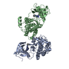
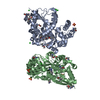
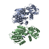
| Title | GLUTAMINE BINDING PROTEIN OPEN LIGAND-FREE STRUCTURE | ||||||
 Components Components | GLUTAMINE BINDING PROTEIN | ||||||
 Keywords Keywords | BINDING PROTEIN / AMINO-ACID TRANSPORT / GLNBP / OPEN FORM | ||||||
| Function / homology |  Function and homology information Function and homology informationL-glutamine binding / L-glutamine import across plasma membrane / glutamine transport / amino acid binding / ligand-gated monoatomic ion channel activity / amino acid transport / ATP-binding cassette (ABC) transporter complex, substrate-binding subunit-containing / outer membrane-bounded periplasmic space / periplasmic space / membrane Similarity search - Function | ||||||
| Biological species |  | ||||||
| Method |  X-RAY DIFFRACTION / ISIRAS / Resolution: 2.3 Å X-RAY DIFFRACTION / ISIRAS / Resolution: 2.3 Å | ||||||
 Authors Authors | Hsiao, C.-D. / Sun, Y.-J. / Rose, J. / Wang, B.-C. | ||||||
 Citation Citation | Journal: J.Mol.Biol. / Year: 1996 Title: The crystal structure of glutamine-binding protein from Escherichia coli. Authors: Hsiao, C.D. / Sun, Y.J. / Rose, J. / Wang, B.C. #1: Journal: J.Mol.Biol. / Year: 1994Title: Crystals of Glutamine-Binding Protein in Various Conformational States Authors: Hsiao, C.D. / Sun, Y.J. / Rose, J. / Cottam, P.F. / Ho, C. / Wang, B.C. #2: Journal: J.Mol.Biol. / Year: 1989Title: Preliminary Crystallographic Analysis of Glutamine-Binding Protein from Escherichia Coli Authors: Chen, P. / Rose, J. / Chung, Y.J. / Wang, B.C. / Shen, Q.C. / Cottam, P.F. / Ho, C. | ||||||
| History |
|
- Structure visualization
| Structure viewer | Molecule: MolmilJmol/JSmol |
|---|
- Downloads & links
-Download
| PDBx/mmCIF format | 1ggg.cif.gz | 98.6 KB | Display | PDBx/mmCIF format |
|---|---|---|---|---|
| PDB format | pdb1ggg.ent.gz | 76.7 KB | Display | PDB format |
| PDBx/mmJSON format | 1ggg.json.gz | Tree view | PDBx/mmJSON format | |
| Others |  Other downloads Other downloads |
-Validation report
| Arichive directory | https://data.pdbj.org/pub/pdb/validation_reports/gg/1gggftp://data.pdbj.org/pub/pdb/validation_reports/gg/1ggg | HTTPS FTP |
|---|
-Related structure data
| Similar structure data |
|---|


-Links
 PDBj
PDBj
- Assembly
| Deposited unit | 
| ||||||||
|---|---|---|---|---|---|---|---|---|---|
| 1 |
| ||||||||
| Unit cell |
|
-Components
| #1: Protein | Mass: 24980.359 Da / Num. of mol.: 2 Source method: isolated from a genetically manipulated source Details: THIS ENTRY REPRESENTS THE OPEN LIGAND FREE STRUCTURE Source: (gene. exp.) #2: Water | ChemComp-HOH / |  Mass: 18.015 Da / Num. of mol.: 159 / Source method: isolated from a natural source / Formula: H2O Mass: 18.015 Da / Num. of mol.: 159 / Source method: isolated from a natural source / Formula: H2OCompound details | THIS ENTRY REPRESENTS THE LIGAND FREE "OPEN" FORM OF THE PROTEIN. UPON BINDING L-GLN, THE PROTEIN ...THIS ENTRY REPRESENTS | |
|---|
-Experimental details
-Experiment
| Experiment | Method: X-RAY DIFFRACTION / Number of used crystals: 1 |
|---|
- Sample preparation
| Crystal | Density Matthews: 3.04 Å3/Da / Density % sol: 53 % | |||||||||||||||||||||||||
|---|---|---|---|---|---|---|---|---|---|---|---|---|---|---|---|---|---|---|---|---|---|---|---|---|---|---|
| Crystal grow | pH: 8.6 / Details: pH 8.6 | |||||||||||||||||||||||||
| Crystal grow | *PLUS Temperature: 18 ℃ / Method: vapor diffusion | |||||||||||||||||||||||||
| Components of the solutions | *PLUS
|
-Data collection
| Diffraction | Mean temperature: 295 K |
|---|---|
| Diffraction source | Source: ROTATING ANODE / Type: RIGAKU RUH3R / Wavelength: 1.5418 |
| Detector | Type: SIEMENS / Detector: AREA DETECTOR / Date: Jul 25, 1990 / Details: SUPPER MIRRORS |
| Radiation | Monochromator: NI FOIL / Monochromatic (M) / Laue (L): M / Scattering type: x-ray |
| Radiation wavelength | Wavelength: 1.5418 Å / Relative weight: 1 |
| Reflection | Resolution: 2.3→48.5 Å / Num. obs: 24247 / % possible obs: 92.6 % / Observed criterion σ(I): -3 / Redundancy: 4.1 % / Rmerge(I) obs: 0.0513 / Net I/σ(I): 17.37 |
| Reflection shell | Resolution: 2.3→2.46 Å / Redundancy: 2.6 % / Rmerge(I) obs: 0.2511 / Mean I/σ(I) obs: 2.045 / % possible all: 61 |
| Reflection | *PLUS Num. measured all: 100231 |
- Processing
| Software |
| ||||||||||||||||||||||||||||||||||||||||||||||||||||||||||||
|---|---|---|---|---|---|---|---|---|---|---|---|---|---|---|---|---|---|---|---|---|---|---|---|---|---|---|---|---|---|---|---|---|---|---|---|---|---|---|---|---|---|---|---|---|---|---|---|---|---|---|---|---|---|---|---|---|---|---|---|---|---|
| Refinement | Method to determine structure: ISIRAS / Resolution: 2.3→10 Å / Cross valid method: THROUGHOUT / σ(F): 2
| ||||||||||||||||||||||||||||||||||||||||||||||||||||||||||||
| Displacement parameters | Biso mean: 24.7 Å2 | ||||||||||||||||||||||||||||||||||||||||||||||||||||||||||||
| Refine analyze | Luzzati coordinate error obs: 0.3 Å | ||||||||||||||||||||||||||||||||||||||||||||||||||||||||||||
| Refinement step | Cycle: LAST / Resolution: 2.3→10 Å
| ||||||||||||||||||||||||||||||||||||||||||||||||||||||||||||
| Refine LS restraints |
| ||||||||||||||||||||||||||||||||||||||||||||||||||||||||||||
| Software | *PLUS Name: X-PLOR / Classification: refinement | ||||||||||||||||||||||||||||||||||||||||||||||||||||||||||||
| Refinement | *PLUS Num. reflection obs: 21735 | ||||||||||||||||||||||||||||||||||||||||||||||||||||||||||||
| Solvent computation | *PLUS | ||||||||||||||||||||||||||||||||||||||||||||||||||||||||||||
| Displacement parameters | *PLUS | ||||||||||||||||||||||||||||||||||||||||||||||||||||||||||||
| LS refinement shell | *PLUS Highest resolution: 2.3 Å / Lowest resolution: 2.4 Å / % reflection Rfree: 0.37 % / Total num. of bins used: 8 / Num. reflection obs: 782 / Rfactor obs: 0.32 |
