 Movie
Movie Controller


+ Open data

- Basic information
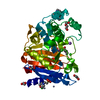
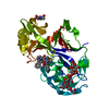
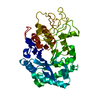
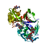
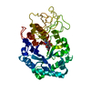
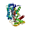
| Entry | Database: PDB / ID: 1g0v | |||||||||
|---|---|---|---|---|---|---|---|---|---|---|
| Title | THE STRUCTURE OF PROTEINASE A COMPLEXED WITH A IA3 MUTANT, MVV | |||||||||
 Components Components |
| |||||||||
 Keywords Keywords | hydrolase/hydrolase inhibitor / Proteinase A / MVV / hydrolase-hydrolase inhibitor COMPLEX | |||||||||
| Function / homology |  Function and homology information Function and homology informationsaccharopepsin / protein catabolic process in the vacuole / microautophagy / cytoplasm to vacuole targeting by the Cvt pathway / oligosaccharide binding / aspartic-type endopeptidase inhibitor activity / pexophagy / fungal-type vacuole / vacuole / endopeptidase inhibitor activity ...saccharopepsin / protein catabolic process in the vacuole / microautophagy / cytoplasm to vacuole targeting by the Cvt pathway / oligosaccharide binding / aspartic-type endopeptidase inhibitor activity / pexophagy / fungal-type vacuole / vacuole / endopeptidase inhibitor activity / : / macroautophagy / autophagy / disordered domain specific binding / peptidase activity / protease binding / aspartic-type endopeptidase activity / endoplasmic reticulum / protein-containing complex / mitochondrion / nucleus / cytoplasm Similarity search - Function | |||||||||
| Biological species |  | |||||||||
| Method |  X-RAY DIFFRACTION / SYNCHROTRON / Resolution: 2 Å X-RAY DIFFRACTION / SYNCHROTRON / Resolution: 2 Å | |||||||||
 Authors Authors | Phylip, L.H. / Lees, W. / Brownsey, B.G. / Bur, D. / Dunn, B.M. / Winther, J. / Gustchina, A. / Li, M. / Copeland, T. / Wlodawer, A. / Kay, J. | |||||||||
 Citation Citation | Journal: J.Biol.Chem. / Year: 2001 Title: The potency and specificity of the interaction between the IA3 inhibitor and its target aspartic proteinase from Saccharomyces cerevisiae. Authors: Phylip, L.H. / Lees, W.E. / Brownsey, B.G. / Bur, D. / Dunn, B.M. / Winther, J.R. / Gustchina, A. / Li, M. / Copeland, T. / Wlodawer, A. / Kay, J. #1: Journal: Nat.Struct.Biol. / Year: 2000Title: The aspartic proteinase from Saccharomyces cerevisiae folds its own inhibitor into helix Authors: Li, M. / Phylip, L.H. / Lees, W. / Winther, J. / Dunn, B. / Wlodawer, A. / Kay, J. / Gustchina, A. | |||||||||
| History |
|
- Structure visualization
| Structure viewer | Molecule: MolmilJmol/JSmol |
|---|
- Downloads & links
-Download
| PDBx/mmCIF format | 1g0v.cif.gz | 92.2 KB | Display | PDBx/mmCIF format |
|---|---|---|---|---|
| PDB format | pdb1g0v.ent.gz | 68.6 KB | Display | PDB format |
| PDBx/mmJSON format | 1g0v.json.gz | Tree view | PDBx/mmJSON format | |
| Others |  Other downloads Other downloads |
-Validation report
| Arichive directory | https://data.pdbj.org/pub/pdb/validation_reports/g0/1g0vftp://data.pdbj.org/pub/pdb/validation_reports/g0/1g0v | HTTPS FTP |
|---|
-Related structure data
| Related structure data | |
|---|---|
| Similar structure data |


-Links
 PDBj
PDBj


- Assembly
| Deposited unit | 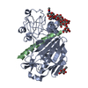
| ||||||||
|---|---|---|---|---|---|---|---|---|---|
| 1 |
| ||||||||
| 2 | 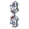
| ||||||||
| 3 | 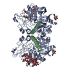
| ||||||||
| Unit cell |
|
-Components
-Protein / Protein/peptide / Non-polymers , 3 types, 231 molecules AB
| #1: Protein | Mass: 35675.422 Da / Num. of mol.: 1 Source method: isolated from a genetically manipulated source Source: (gene. exp.) Production host:  |
|---|---|
| #2: Protein/peptide | Mass: 3493.913 Da / Num. of mol.: 1 / Fragment: MVV, A MUTANT OF IA3 / Mutation: K24M / Source method: obtained synthetically / References: UniProt: P01094 |
| #6: Water | ChemComp-HOH / Mass: 18.015 Da / Num. of mol.: 229 / Source method: isolated from a natural source / Formula: H2O |
-Sugars , 3 types, 3 molecules 

| #3: Polysaccharide | beta-D-mannopyranose-(1-2)-alpha-D-mannopyranose-(1-2)-[alpha-D-mannopyranose-(1-2)-alpha-D- ...beta-D-mannopyranose-(1-2)-alpha-D-mannopyranose-(1-2)-[alpha-D-mannopyranose-(1-2)-alpha-D-mannopyranose-(1-6)]alpha-D-mannopyranose-(1-3)-beta-D-mannopyranose-(1-4)-2-acetamido-2-deoxy-beta-D-glucopyranose-(1-4)-2-acetamido-2-deoxy-beta-D-glucopyranose Source method: isolated from a genetically manipulated source |
|---|---|
| #4: Sugar | ChemComp-MAN /  Type: D-saccharide, alpha linking / Mass: 180.156 Da / Num. of mol.: 1 Type: D-saccharide, alpha linking / Mass: 180.156 Da / Num. of mol.: 1Source method: isolated from a genetically manipulated source Formula: C6H12O6 |
| #5: Sugar | ChemComp-NAG /  Type: D-saccharide, beta linking / Mass: 221.208 Da / Num. of mol.: 1 Type: D-saccharide, beta linking / Mass: 221.208 Da / Num. of mol.: 1Source method: isolated from a genetically manipulated source Formula: C8H15NO6 |
-Details
| Has protein modification | Y |
|---|
-Experimental details
-Experiment
| Experiment | Method: X-RAY DIFFRACTION / Number of used crystals: 1 |
|---|
- Sample preparation
| Crystal | Density Matthews: 3.52 Å3/Da / Density % sol: 65.07 % | |||||||||||||||||||||||||
|---|---|---|---|---|---|---|---|---|---|---|---|---|---|---|---|---|---|---|---|---|---|---|---|---|---|---|
| Crystal grow | Temperature: 298 K / Method: vapor diffusion, hanging drop / pH: 5.6 Details: PEG 1500, (NH4)2SO4, pH 5.6, VAPOR DIFFUSION, HANGING DROP at 298K | |||||||||||||||||||||||||
| Crystal grow | *PLUS pH: 6 / Method: vapor diffusion | |||||||||||||||||||||||||
| Components of the solutions | *PLUS
|
-Data collection
| Diffraction | Mean temperature: 100 K |
|---|---|
| Diffraction source | Source: SYNCHROTRON / Site: NSLS  / Beamline: X9B / Wavelength: 0.92 / Beamline: X9B / Wavelength: 0.92 |
| Detector | Type: ADSC QUANTUM 4 / Detector: CCD / Date: Jul 8, 1999 |
| Radiation | Protocol: SINGLE WAVELENGTH / Monochromatic (M) / Laue (L): M / Scattering type: x-ray |
| Radiation wavelength | Wavelength: 0.92 Å / Relative weight: 1 |
| Reflection | Resolution: 1.9→30 Å / Num. all: 41718 / Num. obs: 41718 / % possible obs: 92.8 % / Observed criterion σ(F): 0 / Observed criterion σ(I): 0 / Redundancy: 5.2 % / Biso Wilson estimate: 7.3 Å2 / Rmerge(I) obs: 0.087 / Net I/σ(I): 12.7 |
| Reflection shell | Resolution: 1.9→1.97 Å / Redundancy: 1.3 % / Rmerge(I) obs: 0.346 / Num. unique all: 3327 / % possible all: 75.5 |
| Reflection | *PLUS Num. measured all: 217446 |
| Reflection shell | *PLUS % possible obs: 75.5 % |
- Processing
| Software |
| |||||||||||||||||||||||||
|---|---|---|---|---|---|---|---|---|---|---|---|---|---|---|---|---|---|---|---|---|---|---|---|---|---|---|
| Refinement | Resolution: 2→24.41 Å / Rfactor Rfree error: 0.005 / Data cutoff high absF: 5393506.27 / Data cutoff low absF: 0 / Isotropic thermal model: RESTRAINED / Cross valid method: THROUGHOUT / σ(F): 0 / σ(I): 0 / Stereochemistry target values: Engh & Huber
| |||||||||||||||||||||||||
| Solvent computation | Solvent model: FLAT MODEL / Bsol: 59.59 Å2 / ksol: 0.362 e/Å3 | |||||||||||||||||||||||||
| Displacement parameters | Biso mean: 20.6 Å2
| |||||||||||||||||||||||||
| Refine analyze |
| |||||||||||||||||||||||||
| Refinement step | Cycle: LAST / Resolution: 2→24.41 Å
| |||||||||||||||||||||||||
| Refine LS restraints |
| |||||||||||||||||||||||||
| LS refinement shell | Resolution: 2→2.13 Å / Rfactor Rfree error: 0.016 / Total num. of bins used: 6
| |||||||||||||||||||||||||
| Xplor file |
| |||||||||||||||||||||||||
| Software | *PLUS Name: CNS / Version: 1 / Classification: refinement | |||||||||||||||||||||||||
| Refine LS restraints | *PLUS
|
