 Movie
Movie Controller

[English] 日本語
 Yorodumi
- PDB-1fbt: THE BISPHOSPHATASE DOMAIN OF THE BIFUNCTIONAL RAT LIVER 6-PHOSPHO... -
+ Open data

- Basic information
| Entry | Database: PDB / ID: 1fbt | ||||||
|---|---|---|---|---|---|---|---|
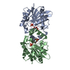
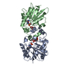
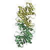
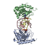
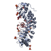
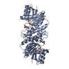
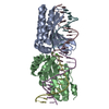
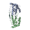
| Title | THE BISPHOSPHATASE DOMAIN OF THE BIFUNCTIONAL RAT LIVER 6-PHOSPHOFRUCTO-2-KINASE/FRUCTOSE-2,6-BISPHOSPHATASE | ||||||
 Components Components | FRUCTOSE-2,6-BISPHOSPHATASE | ||||||
 Keywords Keywords | HYDROLASE / MULTIFUNCTIONAL ENZYME / TRANSFERASE / KINASE | ||||||
| Function / homology |  Function and homology information Function and homology informationpositive regulation of glycolytic process through fructose-6-phosphate / Regulation of glycolysis by fructose 2,6-bisphosphate metabolism / 6-phosphofructo-2-kinase/fructose-2,6-biphosphatase complex / 6-phosphofructo-2-kinase / 6-phosphofructo-2-kinase activity / fructose 2,6-bisphosphate metabolic process / fructose-2,6-bisphosphate 2-phosphatase / fructose-2,6-bisphosphate 2-phosphatase activity / carbohydrate phosphorylation / fructose-6-phosphate binding ...positive regulation of glycolytic process through fructose-6-phosphate / Regulation of glycolysis by fructose 2,6-bisphosphate metabolism / 6-phosphofructo-2-kinase/fructose-2,6-biphosphatase complex / 6-phosphofructo-2-kinase / 6-phosphofructo-2-kinase activity / fructose 2,6-bisphosphate metabolic process / fructose-2,6-bisphosphate 2-phosphatase / fructose-2,6-bisphosphate 2-phosphatase activity / carbohydrate phosphorylation / fructose-6-phosphate binding / fructose metabolic process / response to glucagon / response to starvation / negative regulation of glycolytic process through fructose-6-phosphate / animal organ regeneration / response to cAMP / response to glucocorticoid / response to insulin / kinase binding / ATP binding / identical protein binding / cytosol Similarity search - Function | ||||||
| Biological species |  | ||||||
| Method |  X-RAY DIFFRACTION / SYNCHROTRON / Resolution: 2 Å X-RAY DIFFRACTION / SYNCHROTRON / Resolution: 2 Å | ||||||
 Authors Authors | Lee, Y.-H. / Ogata, C. / Pflugrath, J.W. / Levitt, D.G. / Sarma, R. / Banaszak, L.J. / Pilkis, S.J. | ||||||
 Citation Citation | Journal: Biochemistry / Year: 1996 Title: Crystal structure of the rat liver fructose-2,6-bisphosphatase based on selenomethionine multiwavelength anomalous dispersion phases. Authors: Lee, Y.H. / Ogata, C. / Pflugrath, J.W. / Levitt, D.G. / Sarma, R. / Banaszak, L.J. / Pilkis, S.J. #1: Journal: Annu.Rev.Biochem. / Year: 1995Title: 6-Phosphofructo-2-Kinase/Fructose-2,6-Bisphosphatase: A Metabolic Signaling Enzyme Authors: Pilkis, S.J. / Claus, T.H. / Kurland, I.J. / Lange, A.J. #2: Journal: J.Mol.Biol. / Year: 1994Title: Preliminary X-Ray Analysis of a Truncated Form of Recombinant Fructose-2,6-Bisphosphatase Authors: Lee, Y.H. / Lin, K. / Okar, D. / Alfano, N.L. / Sarma, R. / Pflugrath, J.W. / Pilkis, S.J. | ||||||
| History |
|
- Structure visualization
| Structure viewer | Molecule: MolmilJmol/JSmol |
|---|
- Downloads & links
-Download
| PDBx/mmCIF format | 1fbt.cif.gz | 92.8 KB | Display | PDBx/mmCIF format |
|---|---|---|---|---|
| PDB format | pdb1fbt.ent.gz | 71 KB | Display | PDB format |
| PDBx/mmJSON format | 1fbt.json.gz | Tree view | PDBx/mmJSON format | |
| Others |  Other downloads Other downloads |
-Validation report
| Arichive directory | https://data.pdbj.org/pub/pdb/validation_reports/fb/1fbtftp://data.pdbj.org/pub/pdb/validation_reports/fb/1fbt | HTTPS FTP |
|---|
-Related structure data
| Similar structure data |
|---|


-Links
 PDBj
PDBj


- Assembly
| Deposited unit | 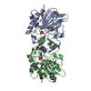
| ||||||||
|---|---|---|---|---|---|---|---|---|---|
| 1 |
| ||||||||
| Unit cell |
| ||||||||
| Noncrystallographic symmetry (NCS) | NCS oper: (Code: given Matrix: (-0.9999, -0.0119, -0.0012), Vector: |
-Components
| #1: Protein | Mass: 22235.746 Da / Num. of mol.: 2 / Mutation: 30 AMINO ACIDS DELETED FROM THE C-TERMINAL Source method: isolated from a genetically manipulated source Source: (gene. exp.) Gene (production host): A CODING REGION WHICH COVERS FRUCTOSE-2,6-BISPHOSPHATASE DOMAIN (RESIDUES 251-440) OF THE RAT LIVER 6-PF-2-K/FRU-2,6-P2ASE (RESIDUES 1-470) Production host:  References: UniProt: P07953, fructose-2,6-bisphosphate 2-phosphatase #2: Chemical |   Mass: 94.971 Da / Num. of mol.: 2 / Source method: obtained synthetically / Formula: PO4 Mass: 94.971 Da / Num. of mol.: 2 / Source method: obtained synthetically / Formula: PO4#3: Water | ChemComp-HOH / |  Mass: 18.015 Da / Num. of mol.: 161 / Source method: isolated from a natural source / Formula: H2O Mass: 18.015 Da / Num. of mol.: 161 / Source method: isolated from a natural source / Formula: H2OCompound details | COMPND MOLECULE: FRU-2,6-BISPHOSPHATASE. THE FRU-2,6-BISPHOSPHATASE IS C-TERMINAL HALF (RESIDUE 251- ...COMPND MOLECULE: FRU-2,6-BISPHOSPHA | Has protein modification | Y | |
|---|
-Experimental details
-Experiment
| Experiment | Method: X-RAY DIFFRACTION / Number of used crystals: 1 |
|---|
- Sample preparation
| Crystal | Density Matthews: 2.88 Å3/Da / Density % sol: 52 % | |||||||||||||||||||||||||||||||||||||||||||||||||||||||
|---|---|---|---|---|---|---|---|---|---|---|---|---|---|---|---|---|---|---|---|---|---|---|---|---|---|---|---|---|---|---|---|---|---|---|---|---|---|---|---|---|---|---|---|---|---|---|---|---|---|---|---|---|---|---|---|---|
| Crystal grow | *PLUS pH: 7 / Method: vapor diffusion, hanging drop | |||||||||||||||||||||||||||||||||||||||||||||||||||||||
| Components of the solutions | *PLUS
|
-Data collection
| Diffraction | Mean temperature: 95 K |
|---|---|
| Diffraction source | Source: SYNCHROTRON / Site: NSLS  / Beamline: X12C / Wavelength: 1.04 / Beamline: X12C / Wavelength: 1.04 |
| Detector | Type: MARRESEARCH / Detector: IMAGE PLATE / Date: Oct 4, 1994 |
| Radiation | Monochromatic (M) / Laue (L): M / Scattering type: x-ray |
| Radiation wavelength | Wavelength: 1.04 Å / Relative weight: 1 |
| Reflection | Num. obs: 32129 / % possible obs: 93.7 % / Observed criterion σ(I): 3 / Redundancy: 6.6 % / Rmerge(I) obs: 0.068 |
| Reflection | *PLUS Highest resolution: 2.5 Å / Lowest resolution: 10 Å / Num. obs: 15137 |
- Processing
| Software |
| ||||||||||||||||||||||||||||||||||||||||||||||||||||||||||||
|---|---|---|---|---|---|---|---|---|---|---|---|---|---|---|---|---|---|---|---|---|---|---|---|---|---|---|---|---|---|---|---|---|---|---|---|---|---|---|---|---|---|---|---|---|---|---|---|---|---|---|---|---|---|---|---|---|---|---|---|---|---|
| Refinement | Resolution: 2→20 Å / σ(F): 2
| ||||||||||||||||||||||||||||||||||||||||||||||||||||||||||||
| Displacement parameters | Biso mean: 27.94 Å2 | ||||||||||||||||||||||||||||||||||||||||||||||||||||||||||||
| Refine analyze | Luzzati coordinate error obs: 0.3 Å | ||||||||||||||||||||||||||||||||||||||||||||||||||||||||||||
| Refinement step | Cycle: LAST / Resolution: 2→20 Å
| ||||||||||||||||||||||||||||||||||||||||||||||||||||||||||||
| Refine LS restraints |
| ||||||||||||||||||||||||||||||||||||||||||||||||||||||||||||
| Software | *PLUS Name: X-PLOR / Classification: refinement | ||||||||||||||||||||||||||||||||||||||||||||||||||||||||||||
| Refinement | *PLUS Highest resolution: 2.5 Å / Lowest resolution: 6 Å / Rfactor obs: 0.208 / Rfactor Rfree: 0.273 | ||||||||||||||||||||||||||||||||||||||||||||||||||||||||||||
| Solvent computation | *PLUS | ||||||||||||||||||||||||||||||||||||||||||||||||||||||||||||
| Displacement parameters | *PLUS Biso mean: 25.6 Å2 | ||||||||||||||||||||||||||||||||||||||||||||||||||||||||||||
| Refine LS restraints | *PLUS
|
