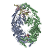
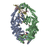
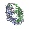
- PDB-1ewq: CRYSTAL STRUCTURE TAQ MUTS COMPLEXED WITH A HETERODUPLEX DNA AT 2... -
+
データを開く
IDまたはキーワード:
読み込み中...
-
基本情報
登録情報
データベース: PDB / ID: 1ewq
タイトル
CRYSTAL STRUCTURE TAQ MUTS COMPLEXED WITH A HETERODUPLEX DNA AT 2.2 A RESOLUTION
要素
DNA (5'-D(*GP*CP*GP*AP*CP*GP*CP*TP*AP*GP*CP*GP*TP*GP*CP*GP*GP*CP*TP*CP*GP*TP*C)-3')
DNA (5'-D(*GP*GP*AP*CP*GP*AP*GP*CP*CP*GP*CP*CP*GP*CP*TP*AP*GP*CP*GP*TP*CP*G)-3')
DNA MISMATCH REPAIR PROTEIN MUTS
キーワード
replication/DNA / Multiple domains of protein / mostly mixed alpha-beta structures / one domain is entirely helical / double stranded helix / replication-DNA COMPLEX
機能・相同性
機能・相同性情報
mismatched DNA binding / ATP-dependent DNA damage sensor activity / mismatch repair / damaged DNA binding / ATP binding 類似検索 - 分子機能
MutS, DNA mismatch repair protein; Chain A, domain 3 / MutS, DNA mismatch repair protein; Chain A, domain 3 - #10 / MutS, connector domain / DNA repair protein MutS, domain I / DNA mismatch repair protein MutS / DNA mismatch repair protein MutS/MSH / DNA mismatch repair protein MutS-like, N-terminal / DNA mismatch repair protein MutS, connector domain / DNA mismatch repair protein MutS, clamp / DNA mismatch repair protein MutS, N-terminal ...MutS, DNA mismatch repair protein; Chain A, domain 3 / MutS, DNA mismatch repair protein; Chain A, domain 3 - #10 / MutS, connector domain / DNA repair protein MutS, domain I / DNA mismatch repair protein MutS / DNA mismatch repair protein MutS/MSH / DNA mismatch repair protein MutS-like, N-terminal / DNA mismatch repair protein MutS, connector domain / DNA mismatch repair protein MutS, clamp / DNA mismatch repair protein MutS, N-terminal / MutS, connector domain superfamily / MutS domain I / MutS domain II / MutS family domain IV / MutS domain III / DNA mismatch repair MutS family / DNA mismatch repair protein MutS, C-terminal / DNA mismatch repair protein MutS, core / DNA mismatch repair protein MutS, core domain superfamily / MutS domain V / DNA mismatch repair proteins mutS family signature. / DNA-binding domain of DNA mismatch repair MUTS family / ATPase domain of DNA mismatch repair MUTS family / MutS, DNA mismatch repair protein, domain I / Nucleotidyltransferase; domain 5 / P-loop containing nucleotide triphosphate hydrolases / Rossmann fold / P-loop containing nucleoside triphosphate hydrolase / 2-Layer Sandwich / Orthogonal Bundle / 3-Layer(aba) Sandwich / Mainly Alpha / Alpha Beta 類似検索 - ドメイン・相同性
DNA / DNA (> 10) / DNA mismatch repair protein MutS 類似検索 - 構成要素
#241 - 2020年1月 20年の分子を振り返って (Twenty Years of Molecules) 類似性 (1)
-
集合体
登録構造単位
C: DNA (5'-D(*GP*CP*GP*AP*CP*GP*CP*TP*AP*GP*CP*GP*TP*GP*CP*GP*GP*CP*TP*CP*GP*TP*C)-3') D: DNA (5'-D(*GP*GP*AP*CP*GP*AP*GP*CP*CP*GP*CP*CP*GP*CP*TP*AP*GP*CP*GP*TP*CP*G)-3') A: DNA MISMATCH REPAIR PROTEIN MUTS B: DNA MISMATCH REPAIR PROTEIN MUTS ヘテロ分子
 ムービー
ムービー コントローラー


 データを開く
 基本情報
基本情報 要素
要素 キーワード
キーワード 機能・相同性情報
 Thermus aquaticus (バクテリア)
 X線回折 / 解像度: 2.2 Å
  データ登録者
 引用
引用 構造の表示
構造の表示 ダウンロードとリンク
 その他のダウンロード
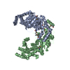


 PDBj
PDBj


 集合体
集合体


 分子量: 96.063 Da / 分子数: 4 / 由来タイプ: 合成 / 式: SO4
 分子量: 62.068 Da / 分子数: 7 / 由来タイプ: 合成 / 式: C2H6O2
 試料調製
試料調製 解析
解析