 Movie
Movie Controller

[English] 日本語
 Yorodumi
- PDB-1ew0: CRYSTAL STRUCTURE ANALYSIS OF THE SENSOR DOMAIN OF RMFIXL(FERROUS... -
+ Open data

- Basic information
| Entry | Database: PDB / ID: 1ew0 | ||||||
|---|---|---|---|---|---|---|---|
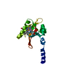
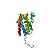
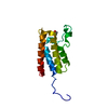
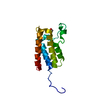
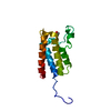
| Title | CRYSTAL STRUCTURE ANALYSIS OF THE SENSOR DOMAIN OF RMFIXL(FERROUS FORM) | ||||||
 Components Components | FIXL | ||||||
 Keywords Keywords |  TRANSFERASE / OXYGEN SENSOR / HEME PROTEIN / HISTIDINE KINASE / RHIZOBIUM MELILOTI TRANSFERASE / OXYGEN SENSOR / HEME PROTEIN / HISTIDINE KINASE / RHIZOBIUM MELILOTI | ||||||
| Function / homology |  Function and homology informationnitrogen fixation / histidine kinase / phosphorelay sensor kinase activity / regulation of DNA-templated transcription / ATP binding / metal ion binding / plasma membrane Function and homology informationnitrogen fixation / histidine kinase / phosphorelay sensor kinase activity / regulation of DNA-templated transcription / ATP binding / metal ion binding / plasma membraneSimilarity search - Function | ||||||
| Biological species |  Sinorhizobium meliloti (bacteria) Sinorhizobium meliloti (bacteria) | ||||||
| Method | X-RAY DIFFRACTION / SYNCHROTRON / Resolution: 1.4 Å | ||||||
 Authors Authors | Miyatake, H. / Mukai, M. / Park, S.-Y. / Adachi, S. / Tamura, K. / Nakamura, H. / Nakamura, K. / Tsuchiya, T. / Iizuka, T. / Shiro, Y. | ||||||
 Citation Citation | Journal: J.MOL.BIOL. / Year: 2000 Title: Sensory mechanism of oxygen sensor FixL from Rhizobium meliloti: crystallographic, mutagenesis and resonance Raman spectroscopic studies Authors: Miyatake, H. / Mukai, M. / Park, S.-Y. / Adachi, S. / Tamura, K. / Nakamura, H. / Nakamura, K. / Tsuchiya, T. / Iizuka, T. / Shiro, Y. #1: Journal: ACTA CRYSTALLOGR.,SECT.D / Year: 1999 Title: Dynamic light-scattering and preliminary crystallographic studies of the sensor domain of the haem-based oxygen sensor FixL from Rhizobium meliloti. Authors: Miyatake, H. / Kanai, M. / Adachi, S. / Nakamura, H. / Tamura, K. / Tanida, H. / Tsuchiya, T. / Iizuka, T. / Shiro, Y. #2: Journal: J.Biol.Chem. / Year: 1999 Title: Iron coordination structure of oxygen sensor FixL characterized by Fe K-edge EXAFS and resonance Raman spectroscopy Authors: Miyatake, H. / Mukai, M. / Adachi, S. / Nakamura, H. / Tamura, K. / Iizuka, T. / Shiro, Y. / Strange, R.W. / Hasnain, S.S. | ||||||
| History |
|
- Structure visualization
| Structure viewer | Molecule: MolmilJmol/JSmol |
|---|
- Downloads & links
-Download
| PDBx/mmCIF format | 1ew0.cif.gz | 41.1 KB | Display | PDBx/mmCIF format |
|---|---|---|---|---|
| PDB format | pdb1ew0.ent.gz | 27.9 KB | Display | PDB format |
| PDBx/mmJSON format | 1ew0.json.gz | Tree view | PDBx/mmJSON format | |
| Others |  Other downloads Other downloads |
-Validation report
| Arichive directory | https://data.pdbj.org/pub/pdb/validation_reports/ew/1ew0ftp://data.pdbj.org/pub/pdb/validation_reports/ew/1ew0 | HTTPS FTP |
|---|
-Related structure data


-Links
 PDBj
PDBj


- Assembly
| Deposited unit | 
| ||||||||
|---|---|---|---|---|---|---|---|---|---|
| 1 |
| ||||||||
| Unit cell |
|
-Components
| #1: Protein | Mass: 14522.385 Da / Num. of mol.: 1 / Fragment: SENSOR DOMAIN Source method: isolated from a genetically manipulated source Source: (gene. exp.) Sinorhizobium meliloti (bacteria) / Plasmid: PET-14B / Production host: Escherichia coli (E. coli)References: UniProt: P10955, Transferases; Transferring phosphorus-containing groups; Phosphotransferases with a nitrogenous group as acceptor |
|---|---|
| #2: Chemical | ChemComp-HEM / Heme B  Mass: 616.487 Da / Num. of mol.: 1 / Source method: obtained synthetically / Formula: C34H32FeN4O4 Mass: 616.487 Da / Num. of mol.: 1 / Source method: obtained synthetically / Formula: C34H32FeN4O4 |
| #3: Water | ChemComp-HOH / Water Mass: 18.015 Da / Num. of mol.: 68 / Source method: isolated from a natural source / Formula: H2O Mass: 18.015 Da / Num. of mol.: 68 / Source method: isolated from a natural source / Formula: H2O |
-Experimental details
-Experiment
| Experiment | Method: X-RAY DIFFRACTION / Number of used crystals: 1 |
|---|
- Sample preparation
| Crystal | Density Matthews: 1.86 Å3/Da / Density % sol: 33.7 % | |||||||||||||||||||||||||
|---|---|---|---|---|---|---|---|---|---|---|---|---|---|---|---|---|---|---|---|---|---|---|---|---|---|---|
| Crystal grow | Temperature: 293 K / Method: vapor diffusion, hanging drop / pH: 4.6 Details: PEG 4000, acetic acid/NaOH, ammonium acetate, pH 4.6, VAPOR DIFFUSION, HANGING DROP, temperature 293K | |||||||||||||||||||||||||
| Crystal grow | *PLUS Temperature: 20 ℃ / pH: 4.5 | |||||||||||||||||||||||||
| Components of the solutions | *PLUS
|
-Data collection
| Diffraction | Mean temperature: 100 K |
|---|---|
| Diffraction source | Source: SYNCHROTRON / Site: SPring-8  / Beamline: BL44B2 / Wavelength: 0.7 / Beamline: BL44B2 / Wavelength: 0.7 |
| Detector | Type: RIGAKU RAXIS IV / Detector: IMAGE PLATE / Date: Jun 10, 1998 |
| Radiation | Protocol: SINGLE WAVELENGTH / Monochromatic (M) / Laue (L): M / Scattering type: x-ray |
| Radiation wavelength | Wavelength: 0.7 Å / Relative weight: 1 |
| Reflection | Resolution: 1.2→100 Å / Num. all: 165005 / Num. obs: 33489 / % possible obs: 92.4 % / Observed criterion σ(F): 0 / Observed criterion σ(I): 0 / Redundancy: 4.93 % / Biso Wilson estimate: 19.2 Å2 / Rmerge(I) obs: 0.051 / Net I/σ(I): 28.7 |
| Reflection shell | Resolution: 1.2→1.28 Å / Redundancy: 3.3 % / Rmerge(I) obs: 0.278 / Num. unique all: 4730 / % possible all: 84.4 |
| Reflection | *PLUS Num. measured all: 165005 |
- Processing
| Software |
| |||||||||||||||||||||||||
|---|---|---|---|---|---|---|---|---|---|---|---|---|---|---|---|---|---|---|---|---|---|---|---|---|---|---|
| Refinement | Resolution: 1.4→20 Å / σ(F): 0 / σ(I): 0 / Stereochemistry target values: Engh & Huber
| |||||||||||||||||||||||||
| Refinement step | Cycle: LAST / Resolution: 1.4→20 Å
| |||||||||||||||||||||||||
| Refine LS restraints |
| |||||||||||||||||||||||||
| Software | *PLUS Name: X-PLOR / Version: 3.851 / Classification: refinement | |||||||||||||||||||||||||
| Refinement | *PLUS Highest resolution: 1.4 Å / Lowest resolution: 20 Å / σ(F): 0 | |||||||||||||||||||||||||
| Solvent computation | *PLUS | |||||||||||||||||||||||||
| Displacement parameters | *PLUS |
