 Movie
Movie Controller


+ Open data

- Basic information
| Entry | Database: PDB / ID: 1eo6 | ||||||
|---|---|---|---|---|---|---|---|
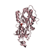
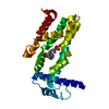
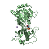
| Title | CRYSTAL STRUCTURE OF GATE-16 | ||||||
 Components Components | GOLGI-ASSOCIATED ATPASE ENHANCER OF 16 KD | ||||||
 Keywords Keywords | PROTEIN BINDING / ubiquitin fold | ||||||
| Function / homology |  Function and homology information Function and homology informationnegative regulation of proteasomal protein catabolic process / autophagy of mitochondrion / protein localization to endoplasmic reticulum / intra-Golgi vesicle-mediated transport / positive regulation of ATP-dependent activity / cellular response to nitrogen starvation / autophagosome membrane / autophagosome assembly / autophagosome / SNARE binding ...negative regulation of proteasomal protein catabolic process / autophagy of mitochondrion / protein localization to endoplasmic reticulum / intra-Golgi vesicle-mediated transport / positive regulation of ATP-dependent activity / cellular response to nitrogen starvation / autophagosome membrane / autophagosome assembly / autophagosome / SNARE binding / macroautophagy / protein transport / ATPase binding / cytoplasmic vesicle / Golgi membrane / ubiquitin protein ligase binding / endoplasmic reticulum membrane / cytosol Similarity search - Function | ||||||
| Biological species |  | ||||||
| Method |  X-RAY DIFFRACTION / SYNCHROTRON / Resolution: 1.8 Å X-RAY DIFFRACTION / SYNCHROTRON / Resolution: 1.8 Å | ||||||
 Authors Authors | Paz, Y. / Elazar, Z. / Fass, D. | ||||||
 Citation Citation | Journal: J.Biol.Chem. / Year: 2000 Title: Structure of GATE-16, membrane transport modulator and mammalian ortholog of autophagocytosis factor Aut7p. Authors: Paz, Y. / Elazar, Z. / Fass, D. #1: Journal: J.Biol.Chem. / Year: 1998Title: Isolation and Characterization of a Novel Low Molecular Weight Protein Involved in Intra-Golgi Traffic Authors: Legesse-Miller, A. / Sagiv, Y. / Porat, A. / Elazar, Z. #2: Journal: Embo J. / Year: 2000Title: GATE-16, a Membrane Transport Modulator Interacts with NSF and the Golgi v-SNARE GOS-28 Authors: Sagiv, Y. / Legesse-Miller, A. / Porat, A. / Elazar, Z. | ||||||
| History |
|
- Structure visualization
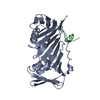
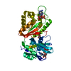
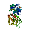
| Structure viewer | Molecule: MolmilJmol/JSmol |
|---|
- Downloads & links
-Download
| PDBx/mmCIF format | 1eo6.cif.gz | 61 KB | Display | PDBx/mmCIF format |
|---|---|---|---|---|
| PDB format | pdb1eo6.ent.gz | 45.5 KB | Display | PDB format |
| PDBx/mmJSON format | 1eo6.json.gz | Tree view | PDBx/mmJSON format | |
| Others |  Other downloads Other downloads |
-Validation report
| Summary document | 1eo6_validation.pdf.gz | 418 KB | Display | wwPDB validaton report |
|---|---|---|---|---|
| Full document | 1eo6_full_validation.pdf.gz | 422.3 KB | Display | |
| Data in XML | 1eo6_validation.xml.gz | 12.8 KB | Display | |
| Data in CIF | 1eo6_validation.cif.gz | 17.7 KB | Display | |
| Arichive directory | https://data.pdbj.org/pub/pdb/validation_reports/eo/1eo6ftp://data.pdbj.org/pub/pdb/validation_reports/eo/1eo6 | HTTPS FTP |
-Related structure data
| Similar structure data |
|---|


-Links
 PDBj
PDBj
- Assembly
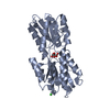
| Deposited unit | 
| ||||||||
|---|---|---|---|---|---|---|---|---|---|
| 1 |
| ||||||||
| Unit cell |
|
-Components
| #1: Protein | Mass: 13686.836 Da / Num. of mol.: 2 Source method: isolated from a genetically manipulated source Source: (gene. exp.)  #2: Water | ChemComp-HOH / |  Mass: 18.015 Da / Num. of mol.: 171 / Source method: isolated from a natural source / Formula: H2O Mass: 18.015 Da / Num. of mol.: 171 / Source method: isolated from a natural source / Formula: H2O |
|---|
-Experimental details
-Experiment
| Experiment | Method: X-RAY DIFFRACTION / Number of used crystals: 1 |
|---|
- Sample preparation
| Crystal | Density Matthews: 2.79 Å3/Da / Density % sol: 55.89 % | ||||||||||||||||||||||||||||||
|---|---|---|---|---|---|---|---|---|---|---|---|---|---|---|---|---|---|---|---|---|---|---|---|---|---|---|---|---|---|---|---|
| Crystal grow | Temperature: 293 K / Method: vapor diffusion, hanging drop / pH: 7.5 Details: PEG 8000, Tris, Calcium acetate, dimethyl sulfoxide, pH 7.5, VAPOR DIFFUSION, HANGING DROP, temperature 293.0K | ||||||||||||||||||||||||||||||
| Crystal grow | *PLUS | ||||||||||||||||||||||||||||||
| Components of the solutions | *PLUS
|
-Data collection
| Diffraction | Mean temperature: 100 K |
|---|---|
| Diffraction source | Source: SYNCHROTRON / Site: ESRF  / Beamline: ID14-2 / Wavelength: 0.933 / Beamline: ID14-2 / Wavelength: 0.933 |
| Detector | Type: ADSC QUANTUM 4 / Detector: CCD / Date: Mar 2, 2000 |
| Radiation | Protocol: SINGLE WAVELENGTH / Monochromatic (M) / Laue (L): M / Scattering type: x-ray |
| Radiation wavelength | Wavelength: 0.933 Å / Relative weight: 1 |
| Reflection | Resolution: 1.8→20 Å / Num. all: 27943 / Num. obs: 27335 / % possible obs: 97.8 % / Observed criterion σ(I): -1.5 / Redundancy: 4.2 % / Biso Wilson estimate: 26.6 Å2 / Rmerge(I) obs: 0.06 / Net I/σ(I): 15.4 |
| Reflection shell | Resolution: 1.8→1.86 Å / Redundancy: 3.9 % / Rmerge(I) obs: 0.252 / Num. unique all: 2777 / % possible all: 97.2 |
| Reflection | *PLUS Highest resolution: 1.8 Å |
| Reflection shell | *PLUS % possible obs: 97.2 % |
- Processing
| Software |
| |||||||||||||||||||||||||
|---|---|---|---|---|---|---|---|---|---|---|---|---|---|---|---|---|---|---|---|---|---|---|---|---|---|---|
| Refinement | Resolution: 1.8→20 Å / σ(F): 0 / Stereochemistry target values: Engh & Huber
| |||||||||||||||||||||||||
| Refinement step | Cycle: LAST / Resolution: 1.8→20 Å
| |||||||||||||||||||||||||
| Refine LS restraints |
| |||||||||||||||||||||||||
| Software | *PLUS Name: CNS / Classification: refinement | |||||||||||||||||||||||||
| Refinement | *PLUS Highest resolution: 1.8 Å / Lowest resolution: 20 Å / σ(F): 0 / Rfactor obs: 0.229 | |||||||||||||||||||||||||
| Solvent computation | *PLUS | |||||||||||||||||||||||||
| Displacement parameters | *PLUS Biso mean: 31.1 Å2 |
