 Movie
Movie Controller

[English] 日本語
 Yorodumi
- PDB-1e85: Cytochrome c' from Alcaligenes xylosoxidans - reduced structure w... -
+ Open data

- Basic information
| Entry | Database: PDB / ID: 1.0E+85 | |||||||||
|---|---|---|---|---|---|---|---|---|---|---|
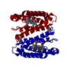
| Title | Cytochrome c' from Alcaligenes xylosoxidans - reduced structure with NO bound to proximal side of heme | |||||||||
 Components Components | CYTOCHROME C' | |||||||||
 Keywords Keywords | ELECTRON TRANSPORT / CYTOCHROME / HEME / 4-HELIX BUNDLE / NITRIC OXIDE | |||||||||
| Function / homology |  Function and homology information Function and homology informationelectron transport chain / electron transfer activity / periplasmic space / iron ion binding / heme binding Similarity search - Function | |||||||||
| Biological species |  ACHROMOBACTER XYLOSOXIDANS (bacteria) ACHROMOBACTER XYLOSOXIDANS (bacteria) | |||||||||
| Method |  X-RAY DIFFRACTION / SYNCHROTRON / MOLECULAR REPLACEMENT / Resolution: 1.35 Å X-RAY DIFFRACTION / SYNCHROTRON / MOLECULAR REPLACEMENT / Resolution: 1.35 Å | |||||||||
 Authors Authors | Lawson, D.M. / Stevenson, C.E.M. / Andrew, C.R. / Eady, R.R. | |||||||||
 Citation Citation | Journal: Embo J. / Year: 2000 Title: Unprecedented Proximal Binding of Nitric Oxide to Heme: Implications for Guanylate Cyclase. Authors: Lawson, D.M. / Stevenson, C.E.M. / Andrew, C.R. / Eady, R.R. #1: Journal: Acta Crystallogr.,Sect.D / Year: 1996Title: The Three-Dimensional Structure of Cytochrome C' from Two Alcaligenes Species and Their Implications for Four-Helix Bundles Authors: Dobbs, A.J. / Faber, H.R. / Anderson, B.F. / Baker, E.N. | |||||||||
| History |
|
- Structure visualization
| Structure viewer | Molecule: MolmilJmol/JSmol |
|---|
- Downloads & links
-Download
| PDBx/mmCIF format | 1e85.cif.gz | 44.6 KB | Display | PDBx/mmCIF format |
|---|---|---|---|---|
| PDB format | pdb1e85.ent.gz | 29.9 KB | Display | PDB format |
| PDBx/mmJSON format | 1e85.json.gz | Tree view | PDBx/mmJSON format | |
| Others |  Other downloads Other downloads |
-Validation report
| Arichive directory | https://data.pdbj.org/pub/pdb/validation_reports/e8/1e85ftp://data.pdbj.org/pub/pdb/validation_reports/e8/1e85 | HTTPS FTP |
|---|
-Related structure data
| Related structure data | 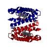 1e83C 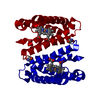 1e84C 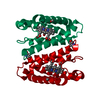 1e86C 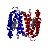 1cgoS S: Starting model for refinement C: citing same article ( |
|---|---|
| Similar structure data |


-Links
 PDBj
PDBj


- Assembly
| Deposited unit | 
| ||||||||
|---|---|---|---|---|---|---|---|---|---|
| 1 | 
| ||||||||
| Unit cell |
| ||||||||
| Components on special symmetry positions |
|
-Components
| #1: Protein | Mass: 13631.442 Da / Num. of mol.: 1 / Source method: isolated from a natural source / Details: FORMERLY KNOWN AS ALCALIGENES SP. / Source: (natural) ACHROMOBACTER XYLOSOXIDANS (bacteria) / Cellular location: PERIPLASM / Strain: NCIB 11015 / References: UniProt: P00138 | ||||
|---|---|---|---|---|---|
| #2: Chemical | ChemComp-HEC /   Mass: 618.503 Da / Num. of mol.: 1 / Source method: obtained synthetically / Formula: C34H34FeN4O4 Mass: 618.503 Da / Num. of mol.: 1 / Source method: obtained synthetically / Formula: C34H34FeN4O4 | ||||
| #3: Chemical | ChemComp-NO /   Mass: 30.006 Da / Num. of mol.: 1 / Source method: obtained synthetically / Formula: NO Mass: 30.006 Da / Num. of mol.: 1 / Source method: obtained synthetically / Formula: NO | ||||
| #4: Water | ChemComp-HOH /  Mass: 18.015 Da / Num. of mol.: 204 / Source method: isolated from a natural source / Formula: H2O Mass: 18.015 Da / Num. of mol.: 204 / Source method: isolated from a natural source / Formula: H2O | ||||
| Compound details | CYTOCHROME| Has protein modification | Y | Sequence details | N-TERMINAL RESIDUE IS PYRROLIDON | |
-Experimental details
-Experiment
| Experiment | Method: X-RAY DIFFRACTION / Number of used crystals: 1 |
|---|
- Sample preparation
| Crystal | Density Matthews: 2.71 Å3/Da / Density % sol: 54.3 % | |||||||||||||||||||||||||
|---|---|---|---|---|---|---|---|---|---|---|---|---|---|---|---|---|---|---|---|---|---|---|---|---|---|---|
| Crystal grow | Method: vapor diffusion, hanging drop / pH: 7.5 Details: HANGING DROP VAPOUR DIFFUSION. PROTEIN AT CONCENTRATION 8 MG/ML WAS MIXED WITH AN EQUAL VOLUME OF WELL SOLUTION CONSISTING OF 55-65% SATURATED AMMONIUM SULFATE IN 100 MM HEPES BUFFER AT PH 7. ...Details: HANGING DROP VAPOUR DIFFUSION. PROTEIN AT CONCENTRATION 8 MG/ML WAS MIXED WITH AN EQUAL VOLUME OF WELL SOLUTION CONSISTING OF 55-65% SATURATED AMMONIUM SULFATE IN 100 MM HEPES BUFFER AT PH 7.5. REDUCED USING MOTHER LIQUOR CONTAINING 20 MM SODIUM DITHIONITE, THEN INCUBATED FOR 6 DAYS IN MOTHER LIQUOR SATURATED WITH NO. | |||||||||||||||||||||||||
| Crystal grow | *PLUS Temperature: 4 ℃ / pH: 7.2 / Method: vapor diffusion, hanging drop | |||||||||||||||||||||||||
| Components of the solutions | *PLUS
|
-Data collection
| Diffraction | Mean temperature: 100 K |
|---|---|
| Diffraction source | Source: SYNCHROTRON / Site: ESRF  / Beamline: ID14-2 / Wavelength: 0.933 / Beamline: ID14-2 / Wavelength: 0.933 |
| Detector | Type: ADSC CCD / Detector: CCD / Date: Jan 15, 2000 / Details: MIRRORS |
| Radiation | Monochromator: DIAMOND / Protocol: SINGLE WAVELENGTH / Monochromatic (M) / Laue (L): M / Scattering type: x-ray |
| Radiation wavelength | Wavelength: 0.933 Å / Relative weight: 1 |
| Reflection | Resolution: 1.35→40 Å / Num. obs: 33925 / % possible obs: 98.8 % / Observed criterion σ(I): -3 / Redundancy: 6.8 % / Biso Wilson estimate: 16 Å2 / Rmerge(I) obs: 0.039 / Net I/σ(I): 39.8 |
| Reflection shell | Resolution: 1.35→1.37 Å / Rmerge(I) obs: 0.227 / Mean I/σ(I) obs: 3.9 / % possible all: 93.1 |
| Reflection shell | *PLUS % possible obs: 93.1 % |
- Processing
| Software |
| ||||||||||||||||||||||||||||||||||||||||||||||||||||||||||||||||||||||||||||||||||||
|---|---|---|---|---|---|---|---|---|---|---|---|---|---|---|---|---|---|---|---|---|---|---|---|---|---|---|---|---|---|---|---|---|---|---|---|---|---|---|---|---|---|---|---|---|---|---|---|---|---|---|---|---|---|---|---|---|---|---|---|---|---|---|---|---|---|---|---|---|---|---|---|---|---|---|---|---|---|---|---|---|---|---|---|---|---|
| Refinement | Method to determine structure: MOLECULAR REPLACEMENT Starting model: PDB ENTRY 1CGO Resolution: 1.35→40 Å / Cross valid method: THROUGHOUT / σ(F): 0 / ESU R: 0.06 / ESU R Free: 0.06 Details: FE ATOM OF HEME REFINED WITH ANISOTROPIC THERMAL PARAMETERS
| ||||||||||||||||||||||||||||||||||||||||||||||||||||||||||||||||||||||||||||||||||||
| Displacement parameters | Biso mean: 18 Å2 | ||||||||||||||||||||||||||||||||||||||||||||||||||||||||||||||||||||||||||||||||||||
| Refinement step | Cycle: LAST / Resolution: 1.35→40 Å
| ||||||||||||||||||||||||||||||||||||||||||||||||||||||||||||||||||||||||||||||||||||
| Refine LS restraints |
| ||||||||||||||||||||||||||||||||||||||||||||||||||||||||||||||||||||||||||||||||||||
| Software | *PLUS Name: REFMAC / Classification: refinement | ||||||||||||||||||||||||||||||||||||||||||||||||||||||||||||||||||||||||||||||||||||
| Refinement | *PLUS Rfactor obs: 0.194 / Rfactor Rfree: 0.22 | ||||||||||||||||||||||||||||||||||||||||||||||||||||||||||||||||||||||||||||||||||||
| Solvent computation | *PLUS | ||||||||||||||||||||||||||||||||||||||||||||||||||||||||||||||||||||||||||||||||||||
| Displacement parameters | *PLUS |
