 Movie
Movie Controller

[English] 日本語
 Yorodumi
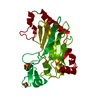
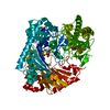
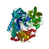
- PDB-1e5h: DELTA-R307A DEACETOXYCEPHALOSPORIN C SYNTHASE COMPLEXED WITH SUCC... -
+ Open data

- Basic information
| Entry | Database: PDB / ID: 1e5h | ||||||
|---|---|---|---|---|---|---|---|
| Title | DELTA-R307A DEACETOXYCEPHALOSPORIN C SYNTHASE COMPLEXED WITH SUCCINATE AND CARBON DIOXIDE | ||||||
 Components Components | DEACETOXYCEPHALOSPORIN C SYNTHASE | ||||||
 Keywords Keywords |  OXIDOREDUCTASE / FERROUS OXYGENASE / CEPHALOSPORIN / 2-OXOGLUTARATE / C-TERMINUS ANTIBIOTICS / OXIDATIVE COUPLING CONTROL OXIDOREDUCTASE / FERROUS OXYGENASE / CEPHALOSPORIN / 2-OXOGLUTARATE / C-TERMINUS ANTIBIOTICS / OXIDATIVE COUPLING CONTROL | ||||||
| Function / homology |  Function and homology informationdeacetoxycephalosporin-C synthase / deacetoxycephalosporin-C synthase activity / L-ascorbic acid binding / antibiotic biosynthetic process / iron ion binding Function and homology informationdeacetoxycephalosporin-C synthase / deacetoxycephalosporin-C synthase activity / L-ascorbic acid binding / antibiotic biosynthetic process / iron ion bindingSimilarity search - Function | ||||||
| Biological species |  STREPTOMYCES CLAVULIGERUS (bacteria) STREPTOMYCES CLAVULIGERUS (bacteria) | ||||||
| Method | X-RAY DIFFRACTION / MOLECULAR REPLACEMENT / Resolution: 1.96 Å | ||||||
 Authors Authors | Lee, H.J. / Lloyd, M.D. / Harlos, K. / Clifton, I.J. / Baldwin, J.E. / Schofield, C.J. | ||||||
 Citation Citation | Journal: J.Mol.Biol. / Year: 2001 Title: Kinetic and Crystallographic Studies on Deacetoxycephalosporin C Synthase (Daocs) Authors: Lee, H.J. / Lloyd, M.D. / Harlos, K. / Clifton, I.J. / Baldwin, J.E. / Schofield, C.J. #1: Journal: J.Mol.Biol. / Year: 1999 Title: Studies on the Active Site of Deacetoxycephalosporin C Synthase (Daocs) Authors: Lloyd, M.D. / Lee, H.J. / Harlos, K. / Zhang, Z.-H. / Baldwin, J.E. / Schofield, C.J. / Charnock, J.M. / Garner, C.D. / Hara, T. / Terrwisscha Van Scheltinga, A.C. / Valegard, K. / Viklund, ...Authors: Lloyd, M.D. / Lee, H.J. / Harlos, K. / Zhang, Z.-H. / Baldwin, J.E. / Schofield, C.J. / Charnock, J.M. / Garner, C.D. / Hara, T. / Terrwisscha Van Scheltinga, A.C. / Valegard, K. / Viklund, J.A.C. / Hajdu, J. / Andersson, I. / Danielsson, A. / Bhikhabhai, R. | ||||||
| History |
|
- Structure visualization
| Structure viewer | Molecule: MolmilJmol/JSmol |
|---|
- Downloads & links
-Download
| PDBx/mmCIF format | 1e5h.cif.gz | 71.5 KB | Display | PDBx/mmCIF format |
|---|---|---|---|---|
| PDB format | pdb1e5h.ent.gz | 51.2 KB | Display | PDB format |
| PDBx/mmJSON format | 1e5h.json.gz | Tree view | PDBx/mmJSON format | |
| Others |  Other downloads Other downloads |
-Validation report
| Arichive directory | https://data.pdbj.org/pub/pdb/validation_reports/e5/1e5hftp://data.pdbj.org/pub/pdb/validation_reports/e5/1e5h | HTTPS FTP |
|---|
-Related structure data
| Related structure data | 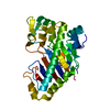 1e5iC  1rxfS S: Starting model for refinement C: citing same article ( |
|---|---|
| Similar structure data |


-Links
 PDBj
PDBj


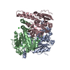
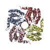
- Assembly
| Deposited unit | 
| ||||||||
|---|---|---|---|---|---|---|---|---|---|
| 1 | 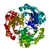
| ||||||||
| Unit cell |
| ||||||||
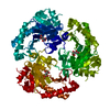
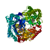
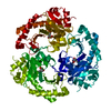
| Details | BIOLOGICALUNIT: ACTIVE AS A MONOMER, TRIMERIC ASSEMBLYOBSERVED IN THE CRYSTAL. |
-Components
| #1: Protein | Mass: 34274.277 Da / Num. of mol.: 1 / Mutation: YES Source method: isolated from a genetically manipulated source Source: (gene. exp.) STREPTOMYCES CLAVULIGERUS (bacteria) / Description: RECOMBINANT E.COLI / Gene: CEFE / Plasmid: PET 24A / Production host: ESCHERICHIA COLI (E. coli) / Strain (production host): BL21(DE3) / References: UniProt: P18548 |
|---|---|
| #2: Chemical | ChemComp-FE2 /   Mass: 55.845 Da / Num. of mol.: 1 / Source method: obtained synthetically / Formula: Fe Mass: 55.845 Da / Num. of mol.: 1 / Source method: obtained synthetically / Formula: Fe |
| #3: Chemical | ChemComp-SIN / Succinic acid  Mass: 118.088 Da / Num. of mol.: 1 / Source method: obtained synthetically / Formula: C4H6O4 Mass: 118.088 Da / Num. of mol.: 1 / Source method: obtained synthetically / Formula: C4H6O4 |
| #4: Chemical | ChemComp-CO2 / Carbon dioxide  Mass: 44.010 Da / Num. of mol.: 1 / Source method: obtained synthetically / Formula: CO2 Mass: 44.010 Da / Num. of mol.: 1 / Source method: obtained synthetically / Formula: CO2 |
| #5: Water | ChemComp-HOH / Water Mass: 18.015 Da / Num. of mol.: 119 / Source method: isolated from a natural source / Formula: H2O Mass: 18.015 Da / Num. of mol.: 119 / Source method: isolated from a natural source / Formula: H2O |
| Compound details | CHAIN A ENGINEERED MUTATION THR308ALA. THE C-TERMINAL RESIDUES SER, LYS, ALA ARE DELETED. FUNCTION: ...CHAIN A ENGINEERED |
| Sequence details | ILE A 50, WRONGLY REPORTED IN DATABASE |
-Experimental details
-Experiment
| Experiment | Method: X-RAY DIFFRACTION / Number of used crystals: 1 |
|---|
- Sample preparation
| Crystal | Density Matthews: 2.39 Å3/Da / Density % sol: 48.16 % | |||||||||||||||||||||||||||||||||||
|---|---|---|---|---|---|---|---|---|---|---|---|---|---|---|---|---|---|---|---|---|---|---|---|---|---|---|---|---|---|---|---|---|---|---|---|---|
| Crystal grow | pH: 7 Details: 100 MM HEPES-NAOH, PH 7.0, 0.5 MM PB(OAC)2(OH2)2, 5 MM NA2SUCCINATE, 5 MM NAHCO3, GLYCEROL 5-10% (W/V), 1.4-1.6 M AMMONIUM SULPHATE | |||||||||||||||||||||||||||||||||||
| Crystal grow | *PLUS Temperature: 20 ℃ / Method: vapor diffusion, hanging drop / Details: Lloyd, M.D., (1999) J.Mol.Biol., 287, 943. | |||||||||||||||||||||||||||||||||||
| Components of the solutions | *PLUS
|
-Data collection
| Diffraction | Mean temperature: 100 K |
|---|---|
| Diffraction source | Source: ROTATING ANODE / Type: RIGAKU / Wavelength: 1.5418 |
| Detector | Type: MAR scanner 300 mm plate / Detector: IMAGE PLATE / Date: Dec 28, 1998 / Details: YALE MIRRORS |
| Radiation | Protocol: SINGLE WAVELENGTH / Monochromatic (M) / Laue (L): M / Scattering type: x-ray |
| Radiation wavelength | Wavelength: 1.5418 Å / Relative weight: 1 |
| Reflection | Resolution: 1.96→30 Å / Num. obs: 21766 / % possible obs: 99.8 % / Redundancy: 5.92 % / Biso Wilson estimate: 21.6 Å2 / Rmerge(I) obs: 0.09 / Net I/σ(I): 25.9 |
| Reflection shell | Resolution: 1.95→1.98 Å / Rmerge(I) obs: 0.281 / Mean I/σ(I) obs: 6.29 / % possible all: 99.6 |
| Reflection | *PLUS Lowest resolution: 30 Å / Num. measured all: 128961 / Rmerge(I) obs: 0.09 |
| Reflection shell | *PLUS % possible obs: 99.6 % |
- Processing
| Software |
| ||||||||||||||||||||||||||||||||||||||||||||||||||||||||||||
|---|---|---|---|---|---|---|---|---|---|---|---|---|---|---|---|---|---|---|---|---|---|---|---|---|---|---|---|---|---|---|---|---|---|---|---|---|---|---|---|---|---|---|---|---|---|---|---|---|---|---|---|---|---|---|---|---|---|---|---|---|---|
| Refinement | Method to determine structure: MOLECULAR REPLACEMENT Starting model: 1RXF Resolution: 1.96→16.57 Å / Rfactor Rfree error: 0.008 / Data cutoff high absF: 2006850.48 / Isotropic thermal model: RESTRAINED / Cross valid method: THROUGHOUT / σ(F): 0 Details: DATA DETWINNED USING CCP4 PROGRAMME DETWIN (A. LESLIE)
| ||||||||||||||||||||||||||||||||||||||||||||||||||||||||||||
| Solvent computation | Solvent model: FLAT MODEL / Bsol: 47.8877 Å2 / ksol: 0.392467 e/Å3 | ||||||||||||||||||||||||||||||||||||||||||||||||||||||||||||
| Displacement parameters | Biso mean: 33.6 Å2
| ||||||||||||||||||||||||||||||||||||||||||||||||||||||||||||
| Refine analyze |
| ||||||||||||||||||||||||||||||||||||||||||||||||||||||||||||
| Refinement step | Cycle: LAST / Resolution: 1.96→16.57 Å
| ||||||||||||||||||||||||||||||||||||||||||||||||||||||||||||
| Refine LS restraints |
| ||||||||||||||||||||||||||||||||||||||||||||||||||||||||||||
| LS refinement shell | Resolution: 1.96→2.08 Å / Rfactor Rfree error: 0.026 / Total num. of bins used: 6
| ||||||||||||||||||||||||||||||||||||||||||||||||||||||||||||
| Xplor file |
| ||||||||||||||||||||||||||||||||||||||||||||||||||||||||||||
| Software | *PLUS Name: CNS / Version: 0.9 / Classification: refinement | ||||||||||||||||||||||||||||||||||||||||||||||||||||||||||||
| Refinement | *PLUS Rfactor obs: 0.22 / Rfactor Rwork: 0.22 | ||||||||||||||||||||||||||||||||||||||||||||||||||||||||||||
| Solvent computation | *PLUS | ||||||||||||||||||||||||||||||||||||||||||||||||||||||||||||
| Displacement parameters | *PLUS | ||||||||||||||||||||||||||||||||||||||||||||||||||||||||||||
| Refine LS restraints | *PLUS
| ||||||||||||||||||||||||||||||||||||||||||||||||||||||||||||
| LS refinement shell | *PLUS Rfactor Rfree: 0.3 |
