 Movie
Movie Controller


+ Open data

- Basic information
| Entry | Database: PDB / ID: 1e2r | ||||||
|---|---|---|---|---|---|---|---|
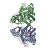
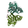
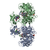
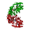
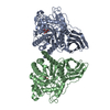
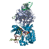
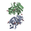
| Title | CYTOCHROME CD1 NITRITE REDUCTASE, REDUCED AND CYANIDE BOUND | ||||||
 Components Components | NITRITE REDUCTASE | ||||||
 Keywords Keywords | OXIDOREDUCTASE / DENITRIFICATION / ELECTRON TRANSPORT / PERIPLASMIC / CYANIDE | ||||||
| Function / homology |  Function and homology information Function and homology informationhydroxylamine reductase / hydroxylamine reductase activity / nitrite reductase (NO-forming) / nitrite reductase (NO-forming) activity / electron transfer activity / periplasmic space / heme binding / metal ion binding Similarity search - Function | ||||||
| Biological species |  PARACOCCUS DENITRIFICANS (bacteria) PARACOCCUS DENITRIFICANS (bacteria) | ||||||
| Method |  X-RAY DIFFRACTION / SYNCHROTRON / MOLECULAR REPLACEMENT / Resolution: 1.59 Å X-RAY DIFFRACTION / SYNCHROTRON / MOLECULAR REPLACEMENT / Resolution: 1.59 Å | ||||||
 Authors Authors | Fulop, V. | ||||||
 Citation Citation | Journal: J.Biol.Chem. / Year: 2000 Title: X-Ray Crystallographic Study of Cyanide Binding Provides Insights Into the Structure-Function Relationship for Cytochrome Cd1 Nitrite Reductase from Paracoccus Pantotrophus. Authors: Jafferji, A. / Allen, J.W. / Ferguson, S.J. / Fulop, V. #1: Journal: J.Mol.Biol. / Year: 1997 Title: Cytochrome Cd1 Structure: Unusual Haem Environments in a Nitrite Reductase and Analysis of Factors Contributing to Beta-Propeller Folds Authors: Baker, S.C. / Saunders, N.F.W. / Willis, A.C. / Ferguson, S.J. / Hajdu, J. / Fulop, V. #2: Journal: Nature / Year: 1997Title: Haem Ligand-Switching During Catalysis in Crystals of a Nitrogen Cycle Enzyme Authors: Williams, P.A. / Fulop, V. / Garman, E.F. / Saunders, N.F.W. / Ferguson, S.J. / Hajdu, J. #3: Journal: Cell(Cambridge,Mass.) / Year: 1995Title: The Anatomy of a Bifunctional Enzyme: Structural Basis for Reduction of Oxygen to Water and Synthesys of Nitric Oxide by Cytochrome Cd1 Authors: Fulop, V. / Moir, J.W.B. / Ferguson, S.J. / Hajdu, J. | ||||||
| History |
|
- Structure visualization
| Structure viewer | Molecule: MolmilJmol/JSmol |
|---|
- Downloads & links
-Download
| PDBx/mmCIF format | 1e2r.cif.gz | 253.6 KB | Display | PDBx/mmCIF format |
|---|---|---|---|---|
| PDB format | pdb1e2r.ent.gz | 202.1 KB | Display | PDB format |
| PDBx/mmJSON format | 1e2r.json.gz | Tree view | PDBx/mmJSON format | |
| Others |  Other downloads Other downloads |
-Validation report
| Arichive directory | https://data.pdbj.org/pub/pdb/validation_reports/e2/1e2rftp://data.pdbj.org/pub/pdb/validation_reports/e2/1e2r | HTTPS FTP |
|---|
-Related structure data
| Related structure data |  1aofS S: Starting model for refinement |
|---|---|
| Similar structure data |


-Links
 PDBj
PDBj


- Assembly
| Deposited unit | 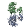
| ||||||||
|---|---|---|---|---|---|---|---|---|---|
| 1 |
| ||||||||
| Unit cell |
| ||||||||
| Noncrystallographic symmetry (NCS) | NCS oper: (Code: given Matrix: (0.721099, 0.545573, 0.427044), Vector: |
-Components
-Protein , 1 types, 2 molecules AB
| #1: Protein | Mass: 62603.633 Da / Num. of mol.: 2 / Source method: isolated from a natural source Details: SUBSP. PARACOCCUS PANTOTROPHUS FORMALLY KNOWN AS THIOSPHAERA PANTOTROPHA Source: (natural) PARACOCCUS DENITRIFICANS (bacteria) / Cellular location: PERIPLASMReferences: UniProt: P72181, nitrite reductase (NO-forming), hydroxylamine reductase |
|---|
-Non-polymers , 5 types, 1351 molecules 


| #2: Chemical |  Mass: 618.503 Da / Num. of mol.: 2 / Source method: obtained synthetically / Formula: C34H34FeN4O4 Mass: 618.503 Da / Num. of mol.: 2 / Source method: obtained synthetically / Formula: C34H34FeN4O4#3: Chemical |  Mass: 712.484 Da / Num. of mol.: 2 / Source method: obtained synthetically / Formula: C34H32FeN4O10 Mass: 712.484 Da / Num. of mol.: 2 / Source method: obtained synthetically / Formula: C34H32FeN4O10#4: Chemical |  Mass: 26.017 Da / Num. of mol.: 2 / Source method: obtained synthetically / Formula: CN Mass: 26.017 Da / Num. of mol.: 2 / Source method: obtained synthetically / Formula: CN#5: Chemical |  Mass: 92.094 Da / Num. of mol.: 2 / Source method: obtained synthetically / Formula: C3H8O3 Mass: 92.094 Da / Num. of mol.: 2 / Source method: obtained synthetically / Formula: C3H8O3#6: Water | ChemComp-HOH / | Mass: 18.015 Da / Num. of mol.: 1343 / Source method: isolated from a natural source / Formula: H2O |
|---|
-Details
| Has protein modification | Y |
|---|
-Experimental details
-Experiment
| Experiment | Method: X-RAY DIFFRACTION / Number of used crystals: 1 |
|---|
- Sample preparation
| Crystal | Density Matthews: 2.41 Å3/Da / Density % sol: 40 % | ||||||||||||||||||||||||
|---|---|---|---|---|---|---|---|---|---|---|---|---|---|---|---|---|---|---|---|---|---|---|---|---|---|
| Crystal grow | pH: 7 Details: 2.3 M AMMONIUM SULFATE, 50MM POTASSIUM PHOSPHATE, PH 7.0, AND CRYOPROTECTANT 15% GLYCEROL | ||||||||||||||||||||||||
| Crystal grow | *PLUS Temperature: 18 ℃ / Method: vapor diffusion, hanging drop | ||||||||||||||||||||||||
| Components of the solutions | *PLUS
|
-Data collection
| Diffraction | Mean temperature: 100 K |
|---|---|
| Diffraction source | Source: SYNCHROTRON / Site: EMBL/DESY, HAMBURG  / Beamline: BW7B / Wavelength: 0.83 / Beamline: BW7B / Wavelength: 0.83 |
| Detector | Type: MARRESEARCH / Detector: IMAGE PLATE / Date: Dec 15, 1998 / Details: MIRRORS |
| Radiation | Protocol: SINGLE WAVELENGTH / Monochromatic (M) / Laue (L): M / Scattering type: x-ray |
| Radiation wavelength | Wavelength: 0.83 Å / Relative weight: 1 |
| Reflection | Resolution: 1.59→25 Å / Num. obs: 156509 / % possible obs: 97.8 % / Observed criterion σ(I): -3 / Redundancy: 3.8 % / Biso Wilson estimate: 16.6 Å2 / Rsym value: 0.081 / Net I/σ(I): 16.8 |
| Reflection shell | Resolution: 1.59→1.65 Å / Redundancy: 2.2 % / Mean I/σ(I) obs: 5.2 / Rsym value: 0.145 / % possible all: 96.7 |
| Reflection | *PLUS Num. measured all: 599187 / Rmerge(I) obs: 0.081 |
- Processing
| Software |
| ||||||||||||||||||||||||||||||||||||||||||||||||||||||||||||
|---|---|---|---|---|---|---|---|---|---|---|---|---|---|---|---|---|---|---|---|---|---|---|---|---|---|---|---|---|---|---|---|---|---|---|---|---|---|---|---|---|---|---|---|---|---|---|---|---|---|---|---|---|---|---|---|---|---|---|---|---|---|
| Refinement | Method to determine structure: MOLECULAR REPLACEMENT Starting model: PDB ENTRY 1AOF Resolution: 1.59→25 Å / Cross valid method: THROUGHOUT / σ(F): 2
| ||||||||||||||||||||||||||||||||||||||||||||||||||||||||||||
| Displacement parameters | Biso mean: 16.1 Å2 | ||||||||||||||||||||||||||||||||||||||||||||||||||||||||||||
| Refine analyze | Luzzati sigma a obs: 0.15 Å | ||||||||||||||||||||||||||||||||||||||||||||||||||||||||||||
| Refinement step | Cycle: LAST / Resolution: 1.59→25 Å
| ||||||||||||||||||||||||||||||||||||||||||||||||||||||||||||
| Refine LS restraints |
| ||||||||||||||||||||||||||||||||||||||||||||||||||||||||||||
| LS refinement shell | Resolution: 1.59→1.62 Å / Total num. of bins used: 20
| ||||||||||||||||||||||||||||||||||||||||||||||||||||||||||||
| Software | *PLUS Name: X-PLOR / Version: 3.851 / Classification: refinement | ||||||||||||||||||||||||||||||||||||||||||||||||||||||||||||
| Refine LS restraints | *PLUS
| ||||||||||||||||||||||||||||||||||||||||||||||||||||||||||||
| LS refinement shell | *PLUS Rfactor Rfree: 0.23 |
