 ムービー
ムービー コントローラー


+ データを開く

- 基本情報
基本情報
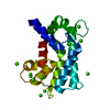
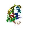
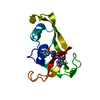
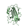
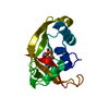
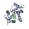
| 登録情報 | データベース: PDB / ID: 1dyt | |||||||||
|---|---|---|---|---|---|---|---|---|---|---|
| タイトル | X-ray crystal structure of ECP (RNase 3) at 1.75 A | |||||||||
 要素 要素 | EOSINOPHIL CATIONIC PROTEIN | |||||||||
 キーワード キーワード | HYDROLASE / EOSINOPHIL CATIONIC PROTEIN / ECP / RNASE 3 | |||||||||
| 機能・相同性 |  機能・相同性情報 機能・相同性情報induction of bacterial agglutination / 加水分解酵素; エステル加水分解酵素; 3'-リン酸モノエステル産生エンドリボヌクレアーゼ / RNA catabolic process / Antimicrobial peptides / RNA nuclease activity / innate immune response in mucosa / lipopolysaccharide binding / chemotaxis / azurophil granule lumen / antimicrobial humoral immune response mediated by antimicrobial peptide ...induction of bacterial agglutination / 加水分解酵素; エステル加水分解酵素; 3'-リン酸モノエステル産生エンドリボヌクレアーゼ / RNA catabolic process / Antimicrobial peptides / RNA nuclease activity / innate immune response in mucosa / lipopolysaccharide binding / chemotaxis / azurophil granule lumen / antimicrobial humoral immune response mediated by antimicrobial peptide / antibacterial humoral response / endonuclease activity / nucleic acid binding / defense response to Gram-negative bacterium / defense response to Gram-positive bacterium / innate immune response / Neutrophil degranulation / extracellular space / extracellular region 類似検索 - 分子機能 | |||||||||
| 生物種 |  HOMO SAPIENS (ヒト) HOMO SAPIENS (ヒト) | |||||||||
| 手法 |  X線回折 / シンクロトロン / 分子置換 / 解像度: 1.75 Å X線回折 / シンクロトロン / 分子置換 / 解像度: 1.75 Å | |||||||||
 データ登録者 データ登録者 | Mallorqui-Fernandez, G. / Pous, J. / Peracaula, R. / Maeda, T. / Tada, H. / Yamada, H. / Seno, M. / De Llorens, R. / Gomis-Rueth, F.X. / Coll, M. | |||||||||
 引用 引用 | ジャーナル: J.Mol.Biol. / 年: 2000 タイトル: Three-Dimensional Crystal Structure of Human Eosinophil Cationic Protein (Rnase 3) at 1.75 A Resolution. 著者: Mallorqui-Fernandez, G. / Pous, J. / Peracaula, R. / Maeda, T. / Tada, H. / Yamada, H. / Seno, M. / De Llorens, R. / Gomis-Rueth, F.X. / Coll, M. | |||||||||
| 履歴 |
|
- 構造の表示
構造の表示
| 構造ビューア | 分子: MolmilJmol/JSmol |
|---|
- ダウンロードとリンク
-ダウンロード
| PDBx/mmCIF形式 | 1dyt.cif.gz | 76.2 KB | 表示 | PDBx/mmCIF形式 |
|---|---|---|---|---|
| PDB形式 | pdb1dyt.ent.gz | 57.9 KB | 表示 | PDB形式 |
| PDBx/mmJSON形式 | 1dyt.json.gz | ツリー表示 | PDBx/mmJSON形式 | |
| その他 |  その他のダウンロード その他のダウンロード |
-検証レポート
| アーカイブディレクトリ | https://data.pdbj.org/pub/pdb/validation_reports/dy/1dytftp://data.pdbj.org/pub/pdb/validation_reports/dy/1dyt | HTTPS FTP |
|---|
-関連構造データ


-リンク
 PDBj
PDBj


- 集合体
集合体
| 登録構造単位 | 
| ||||||||||||
|---|---|---|---|---|---|---|---|---|---|---|---|---|---|
| 1 | 
| ||||||||||||
| 2 | 
| ||||||||||||
| 単位格子 |
| ||||||||||||
| Components on special symmetry positions |
|
-要素
| #1: タンパク質 | 分子量: 15598.876 Da / 分子数: 2 / 由来タイプ: 組換発現 / 由来: (組換発現) HOMO SAPIENS (ヒト) / Cell: EOSINOPHIL / 遺伝子: RNASE3 / 発現宿主:  参照: UniProt: P12724, 加水分解酵素; エステル加水分解酵素; 3'-リン酸モノエステル産生エンドリボヌクレアーゼ #2: 化合物 |   分子量: 55.845 Da / 分子数: 2 / 由来タイプ: 合成 / 式: Fe 分子量: 55.845 Da / 分子数: 2 / 由来タイプ: 合成 / 式: Fe#3: 化合物 | ChemComp-CIT /   分子量: 192.124 Da / 分子数: 4 / 由来タイプ: 合成 / 式: C6H8O7 分子量: 192.124 Da / 分子数: 4 / 由来タイプ: 合成 / 式: C6H8O7#4: 水 | ChemComp-HOH / |  分子量: 18.015 Da / 分子数: 332 / 由来タイプ: 天然 / 式: H2O 分子量: 18.015 Da / 分子数: 332 / 由来タイプ: 天然 / 式: H2OHas protein modification | Y | |
|---|
-実験情報
-実験
| 実験 | 手法: X線回折 / 使用した結晶の数: 1 |
|---|
- 試料調製
試料調製
| 結晶 | マシュー密度: 2.8 Å3/Da / 溶媒含有率: 50 % | ||||||||||||||||||||||||||||||
|---|---|---|---|---|---|---|---|---|---|---|---|---|---|---|---|---|---|---|---|---|---|---|---|---|---|---|---|---|---|---|---|
| 結晶化 | pH: 5.2 詳細: 8% JEFFAMINE M-600, 0.1M SODIUM CITRATE PH 5.2, 0.01M IRON CHLORIDE | ||||||||||||||||||||||||||||||
| 結晶 | *PLUS 溶媒含有率: 50 % | ||||||||||||||||||||||||||||||
| 結晶化 | *PLUS 手法: 蒸気拡散法, ハンギングドロップ法 | ||||||||||||||||||||||||||||||
| 溶液の組成 | *PLUS
|
-データ収集
| 回折 | 平均測定温度: 100 K |
|---|---|
| 放射光源 | 由来: シンクロトロン / サイト: ELETTRA  / ビームライン: 5.2R / 波長: 1.05271 / ビームライン: 5.2R / 波長: 1.05271 |
| 検出器 | タイプ: MARRESEARCH / 検出器: IMAGE PLATE / 日付: 1999年3月15日 / 詳細: MIRRORS |
| 放射 | モノクロメーター: TOROIDAL MIRROR / プロトコル: SINGLE WAVELENGTH / 単色(M)・ラウエ(L): M / 散乱光タイプ: x-ray |
| 放射波長 | 波長: 1.05271 Å / 相対比: 1 |
| 反射 | 解像度: 1.75→22 Å / Num. obs: 35401 / % possible obs: 99.5 % / Observed criterion σ(I): -3 / 冗長度: 1.9 % / Rmerge(I) obs: 0.033 / Rsym value: 0.033 / Net I/σ(I): 16.3 |
| 反射 シェル | 解像度: 1.75→1.81 Å / 冗長度: 2.1 % / Rmerge(I) obs: 0.03 / Mean I/σ(I) obs: 1.75 / Rsym value: 0.03 / % possible all: 99.9 |
| 反射 | *PLUS Num. measured all: 58607 |
| 反射 シェル | *PLUS % possible obs: 99.9 % / Rmerge(I) obs: 0.03 |
- 解析
解析
| ソフトウェア |
| ||||||||||||||||||||||||||||||||||||||||||||||||||||||||||||
|---|---|---|---|---|---|---|---|---|---|---|---|---|---|---|---|---|---|---|---|---|---|---|---|---|---|---|---|---|---|---|---|---|---|---|---|---|---|---|---|---|---|---|---|---|---|---|---|---|---|---|---|---|---|---|---|---|---|---|---|---|---|
| 精密化 | 構造決定の手法: 分子置換 開始モデル: EOSINOPHIL DERIVED PROTEIN 解像度: 1.75→20 Å / Rfactor Rfree error: 8.0E-5 / Data cutoff high absF: 10000 / 交差検証法: MLF / σ(F): 0
| ||||||||||||||||||||||||||||||||||||||||||||||||||||||||||||
| 溶媒の処理 | 溶媒モデル: DENSITY MODIFICATION | ||||||||||||||||||||||||||||||||||||||||||||||||||||||||||||
| 原子変位パラメータ | Biso mean: 27.403 Å2 | ||||||||||||||||||||||||||||||||||||||||||||||||||||||||||||
| 精密化ステップ | サイクル: LAST / 解像度: 1.75→20 Å
| ||||||||||||||||||||||||||||||||||||||||||||||||||||||||||||
| 拘束条件 |
| ||||||||||||||||||||||||||||||||||||||||||||||||||||||||||||
| Xplor file |
| ||||||||||||||||||||||||||||||||||||||||||||||||||||||||||||
| ソフトウェア | *PLUS 名称: CNS / バージョン: 0.9A / 分類: refinement | ||||||||||||||||||||||||||||||||||||||||||||||||||||||||||||
| 精密化 | *PLUS 最低解像度: 20 Å | ||||||||||||||||||||||||||||||||||||||||||||||||||||||||||||
| 溶媒の処理 | *PLUS | ||||||||||||||||||||||||||||||||||||||||||||||||||||||||||||
| 原子変位パラメータ | *PLUS |
