 Movie
Movie Controller

[English] 日本語
 Yorodumi
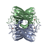
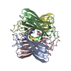
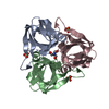
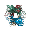
- PDB-1dqk: CRYSTAL STRUCTURE OF SUPEROXIDE REDUCTASE IN THE REDUCED STATE AT... -
+ Open data

- Basic information
| Entry | Database: PDB / ID: 1dqk | ||||||
|---|---|---|---|---|---|---|---|
| Title | CRYSTAL STRUCTURE OF SUPEROXIDE REDUCTASE IN THE REDUCED STATE AT 2.0 ANGSTROMS RESOLUTION | ||||||
 Components Components | SUPEROXIDE REDUCTASE | ||||||
 Keywords Keywords | OXIDOREDUCTASE / non-heme mononuclear iron protein / immunoglobulin-like (Ig) beta barrel fold | ||||||
| Function / homology |  Function and homology information Function and homology information | ||||||
| Biological species |   Pyrococcus furiosus (archaea) Pyrococcus furiosus (archaea) | ||||||
| Method | X-RAY DIFFRACTION / Resolution: 2 Å | ||||||
 Authors Authors | Yeh, A.P. / Hu, Y. / Jenney Jr., F.E. / Adams, M.W.W. / Rees, D.C. | ||||||
 Citation Citation | Journal: Biochemistry / Year: 2000 Title: Structures of the superoxide reductase from Pyrococcus furiosus in the oxidized and reduced states. Authors: Yeh, A.P. / Hu, Y. / Jenney Jr., F.E. / Adams, M.W. / Rees, D.C. #1: Journal: Science / Year: 1999Title: Anaerobic Microbes: Oxygen Detoxification Without Superoxide Dismutase Authors: Jenney Jr., F.E. / Verhagen, M.F. / Cui, X. / Adams, M.W.W. | ||||||
| History |
|
- Structure visualization
| Structure viewer | Molecule: MolmilJmol/JSmol |
|---|
- Downloads & links
-Download
| PDBx/mmCIF format | 1dqk.cif.gz | 117 KB | Display | PDBx/mmCIF format |
|---|---|---|---|---|
| PDB format | pdb1dqk.ent.gz | 91.9 KB | Display | PDB format |
| PDBx/mmJSON format | 1dqk.json.gz | Tree view | PDBx/mmJSON format | |
| Others |  Other downloads Other downloads |
-Validation report
| Arichive directory | https://data.pdbj.org/pub/pdb/validation_reports/dq/1dqkftp://data.pdbj.org/pub/pdb/validation_reports/dq/1dqk | HTTPS FTP |
|---|
-Related structure data


-Links
 PDBj
PDBj


- Assembly
| Deposited unit | 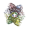
| ||||||||
|---|---|---|---|---|---|---|---|---|---|
| 1 |
| ||||||||
| Unit cell |
|
-Components
| #1: Protein | / SOR Mass: 14344.189 Da / Num. of mol.: 4 / Source method: isolated from a natural source / Source: (natural) Pyrococcus furiosus (archaea) / References: UniProt: P82385#2: Chemical | ChemComp-FE2 /   Mass: 55.845 Da / Num. of mol.: 4 / Source method: obtained synthetically / Formula: Fe Mass: 55.845 Da / Num. of mol.: 4 / Source method: obtained synthetically / Formula: Fe#3: Water | ChemComp-HOH / | Water Mass: 18.015 Da / Num. of mol.: 330 / Source method: isolated from a natural source / Formula: H2O Mass: 18.015 Da / Num. of mol.: 330 / Source method: isolated from a natural source / Formula: H2O |
|---|
-Experimental details
-Experiment
| Experiment | Method: X-RAY DIFFRACTION / Number of used crystals: 1 |
|---|
- Sample preparation
| Crystal | Density Matthews: 2.02 Å3/Da / Density % sol: 39.19 % | ||||||||||||||||||||||||||||||||||||||||||||||||
|---|---|---|---|---|---|---|---|---|---|---|---|---|---|---|---|---|---|---|---|---|---|---|---|---|---|---|---|---|---|---|---|---|---|---|---|---|---|---|---|---|---|---|---|---|---|---|---|---|---|
| Crystal grow | Temperature: 295 K / Method: vapor diffusion, sitting drop / pH: 8 Details: PEG4000, Tris-hydrochloride, pH 8.0, glycerol, sodium chloride, ethanol, VAPOR DIFFUSION, SITTING DROP, temperature 295K | ||||||||||||||||||||||||||||||||||||||||||||||||
| Crystal | *PLUS Density % sol: 44 % | ||||||||||||||||||||||||||||||||||||||||||||||||
| Crystal grow | *PLUS | ||||||||||||||||||||||||||||||||||||||||||||||||
| Components of the solutions | *PLUS
|
-Data collection
| Diffraction | Mean temperature: 90 K |
|---|---|
| Diffraction source | Source: ROTATING ANODE / Type: RIGAKU RU200 / Wavelength: 1.5418 |
| Detector | Type: RIGAKU RAXIS IV / Detector: IMAGE PLATE / Date: May 1, 1998 |
| Radiation | Protocol: SINGLE WAVELENGTH / Monochromatic (M) / Laue (L): M / Scattering type: x-ray |
| Radiation wavelength | Wavelength: 1.5418 Å / Relative weight: 1 |
| Reflection | Resolution: 2→22.7 Å / Num. all: 31825 / Num. obs: 31825 / % possible obs: 98.7 % / Observed criterion σ(F): 0 / Observed criterion σ(I): 0 / Redundancy: 2.9 % / Biso Wilson estimate: 25.9 Å2 / Rmerge(I) obs: 0.06 / Net I/σ(I): 19.2 |
| Reflection shell | Resolution: 2→2.07 Å / Redundancy: 2.5 % / Rmerge(I) obs: 0.242 / Num. unique all: 3038 / % possible all: 95.7 |
| Reflection | *PLUS Num. measured all: 92775 / Rmerge(I) obs: 0.06 |
| Reflection shell | *PLUS % possible obs: 95.7 % / Mean I/σ(I) obs: 4.7 |
- Processing
| Software |
| |||||||||||||||||||||||||
|---|---|---|---|---|---|---|---|---|---|---|---|---|---|---|---|---|---|---|---|---|---|---|---|---|---|---|
| Refinement | Resolution: 2→22.7 Å / σ(F): 0 / σ(I): 0 / Stereochemistry target values: Engh & Huber Details: Since SOR contains iron and the data set was collected at a wavelength near the iron edge, an anomalous signal was expected. Therefore, the structure was refined against the separate Friedel mates
| |||||||||||||||||||||||||
| Refinement step | Cycle: LAST / Resolution: 2→22.7 Å
| |||||||||||||||||||||||||
| Refine LS restraints |
| |||||||||||||||||||||||||
| Software | *PLUS Name: X-PLOR / Version: 3.851 / Classification: refinement | |||||||||||||||||||||||||
| Refinement | *PLUS Highest resolution: 2 Å / Lowest resolution: 22.7 Å / σ(F): 0 / % reflection Rfree: 3 % / Rfactor obs: 0.222 | |||||||||||||||||||||||||
| Solvent computation | *PLUS | |||||||||||||||||||||||||
| Displacement parameters | *PLUS | |||||||||||||||||||||||||
| Refine LS restraints | *PLUS
|
