 Movie
Movie Controller

[English] 日本語
 Yorodumi
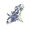
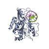
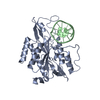
- PDB-1cl8: A PRE-TRANSITION STATE ECO RI ENDONUCLEASE/COGNATE DNA (TCGCGAPTT... -
+ Open data

- Basic information
| Entry | Database: PDB / ID: 1cl8 | ||||||
|---|---|---|---|---|---|---|---|
| Title | A PRE-TRANSITION STATE ECO RI ENDONUCLEASE/COGNATE DNA (TCGCGAPTTCGCG) COMPLEX WITH DNA BASE ANALOG PURINE (P) | ||||||
 Components Components |
| ||||||
 Keywords Keywords | PROTEIN/DNA / ENDONUCLEASE/DNA / DNA BASE ANALOG /  PROTEIN-DNA COMPLEX PROTEIN-DNA COMPLEX | ||||||
| Function / homology |  Function and homology informationtype II site-specific deoxyribonuclease / type II site-specific deoxyribonuclease activity / DNA restriction-modification system / magnesium ion binding / DNA binding Function and homology informationtype II site-specific deoxyribonuclease / type II site-specific deoxyribonuclease activity / DNA restriction-modification system / magnesium ion binding / DNA bindingSimilarity search - Function | ||||||
| Biological species |  Escherichia coli (E. coli) Escherichia coli (E. coli) | ||||||
| Method | X-RAY DIFFRACTION / SYNCHROTRON / OTHER / Resolution: 1.8 Å | ||||||
 Authors Authors | Horvath, M. / Rosenberg, J.M. | ||||||
 Citation Citation | Journal: To be Published Title: Iap (Inner-Adenine to Purine): A Cognate EcoRI-DNA Base Analog Complex Authors: Horvath, M. / Rosenberg, J.M. | ||||||
| History |
|
- Structure visualization
| Structure viewer | Molecule: MolmilJmol/JSmol |
|---|
- Downloads & links
-Download
| PDBx/mmCIF format | 1cl8.cif.gz | 78.1 KB | Display | PDBx/mmCIF format |
|---|---|---|---|---|
| PDB format | pdb1cl8.ent.gz | 54.1 KB | Display | PDB format |
| PDBx/mmJSON format | 1cl8.json.gz | Tree view | PDBx/mmJSON format | |
| Others |  Other downloads Other downloads |
-Validation report
| Arichive directory | https://data.pdbj.org/pub/pdb/validation_reports/cl/1cl8ftp://data.pdbj.org/pub/pdb/validation_reports/cl/1cl8 | HTTPS FTP |
|---|
-Related structure data
| Related structure data |  1ckqS S: Starting model for refinement |
|---|---|
| Similar structure data |


-Links
 PDBj
PDBj


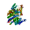
- Assembly
| Deposited unit | 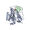
| ||||||||||
|---|---|---|---|---|---|---|---|---|---|---|---|
| 1 | 
| ||||||||||
| Unit cell |
|
-Components
| #1: DNA chain | Mass: 3952.570 Da / Num. of mol.: 1 / Source method: obtained synthetically |
|---|---|
| #2: Protein | Mass: 30970.184 Da / Num. of mol.: 1 / Source method: isolated from a natural source / Source: (natural) Escherichia coli (E. coli) / References: UniProt: P00642 |
| #3: Water | ChemComp-HOH / Water Mass: 18.015 Da / Num. of mol.: 155 / Source method: isolated from a natural source / Formula: H2O Mass: 18.015 Da / Num. of mol.: 155 / Source method: isolated from a natural source / Formula: H2O |
| Nonpolymer details | PRN IS ADENINE WITH THE N6 AMINO GROUP REMOVED |
-Experimental details
-Experiment
| Experiment | Method: X-RAY DIFFRACTION / Number of used crystals: 1 |
|---|
- Sample preparation
| Crystal | Density Matthews: 2.8 Å3/Da / Density % sol: 58 % |
|---|---|
| Crystal grow | Method: vapor diffusion, hanging drop / pH: 7.5 / Details: pH 7.5, VAPOR DIFFUSION, HANGING DROP |
-Data collection
| Diffraction | Mean temperature: 100 K |
|---|---|
| Diffraction source | Source: SYNCHROTRON / Site: CHESS  / Beamline: F1 / Wavelength: 1.1 / Beamline: F1 / Wavelength: 1.1 |
| Detector | Detector: IMAGE PLATE / Date: Apr 15, 1998 / Details: MIRRORS |
| Radiation | Protocol: SINGLE WAVELENGTH / Monochromatic (M) / Laue (L): M / Scattering type: x-ray |
| Radiation wavelength | Wavelength: 1.1 Å / Relative weight: 1 |
| Reflection | Resolution: 1.8→8 Å / Num. obs: 35192 / % possible obs: 98.3 % / Observed criterion σ(I): 2 / Redundancy: 10.3 % / Rmerge(I) obs: 0.105 / Net I/σ(I): 19.3 |
| Reflection shell | Resolution: 1.8→1.85 Å / Redundancy: 4 % / Rmerge(I) obs: 0.432 / Mean I/σ(I) obs: 2.6 / % possible all: 91 |
- Processing
| Software |
| ||||||||||||||||||||||||||||||||||||||||||||||||||||||||||||
|---|---|---|---|---|---|---|---|---|---|---|---|---|---|---|---|---|---|---|---|---|---|---|---|---|---|---|---|---|---|---|---|---|---|---|---|---|---|---|---|---|---|---|---|---|---|---|---|---|---|---|---|---|---|---|---|---|---|---|---|---|---|
| Refinement | Method to determine structure: OTHER Starting model: PDB ENTRY 1CKQ Resolution: 1.8→8 Å / Data cutoff high absF: 10000000 / Data cutoff low absF: 0.001 / Isotropic thermal model: RESTRAINED / Cross valid method: RFREE / σ(F): 2 Details: DNA-RNA.PARAM WAS MODIFIED TO CONTAIN A MODIFIED RESIDUE PRN
| ||||||||||||||||||||||||||||||||||||||||||||||||||||||||||||
| Displacement parameters | Biso mean: 36.1 Å2 | ||||||||||||||||||||||||||||||||||||||||||||||||||||||||||||
| Refine analyze |
| ||||||||||||||||||||||||||||||||||||||||||||||||||||||||||||
| Refinement step | Cycle: LAST / Resolution: 1.8→8 Å
| ||||||||||||||||||||||||||||||||||||||||||||||||||||||||||||
| Refine LS restraints |
| ||||||||||||||||||||||||||||||||||||||||||||||||||||||||||||
| LS refinement shell | Resolution: 1.8→1.88 Å / Total num. of bins used: 8
| ||||||||||||||||||||||||||||||||||||||||||||||||||||||||||||
| Xplor file |
|
