 Movie
Movie Controller

[English] 日本語
 Yorodumi
- PDB-1bwr: PROBING THE SUBSTRATE SPECIFICITY OF THE INTRACELLULAR BRAIN PLAT... -
+ Open data

- Basic information
| Entry | Database: PDB / ID: 1bwr | ||||||
|---|---|---|---|---|---|---|---|
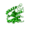
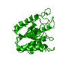
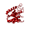
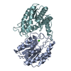
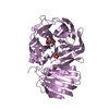
| Title | PROBING THE SUBSTRATE SPECIFICITY OF THE INTRACELLULAR BRAIN PLATELET-ACTIVATING FACTOR ACETYLHYDROLASE | ||||||
 Components Components | PLATELET-ACTIVATING FACTOR ACETYLHYDROLASE | ||||||
 Keywords Keywords | HYDROLASE / ACETYLHYDROLASE | ||||||
| Function / homology |  Function and homology information Function and homology informationplatelet-activating factor acetyltransferase activity / 1-alkyl-2-acetylglycerophosphocholine esterase / 1-alkyl-2-acetylglycerophosphocholine esterase complex / COPI-independent Golgi-to-ER retrograde traffic / 1-alkyl-2-acetylglycerophosphocholine esterase activity / lipid catabolic process / spermatogenesis / protein heterodimerization activity / protein homodimerization activity / cytoplasm Similarity search - Function | ||||||
| Biological species |  | ||||||
| Method |  X-RAY DIFFRACTION / MOLECULAR REPLACEMENT / Resolution: 2.4 Å X-RAY DIFFRACTION / MOLECULAR REPLACEMENT / Resolution: 2.4 Å | ||||||
 Authors Authors | Ho, Y.S. / Sheffield, P.J. / Masuyama, J. / Arai, H. / Li, J. / Aoki, J. / Inoue, K. / Derewenda, U. / Derewenda, Z. | ||||||
 Citation Citation | Journal: Protein Eng. / Year: 1999 Title: Probing the Substrate Specificity of the Intracellular Brain Platelet-Activating Factor Acetylhydrolase Authors: Ho, Y.S. / Sheffield, P.J. / Masuyama, J. / Arai, H. / Li, J. / Aoki, J. / Inoue, K. / Derewenda, U. / Derewenda, Z.S. #1: Journal: Nature / Year: 1997Title: Brain Acetylhydrolase that Inactivates Platelet-Activating Factor is a G-Protein-Like Trimer Authors: Ho, Y.S. / Swenson, L. / Derewenda, U. / Serre, L. / Wei, Y. / Dauter, Z. / Hattori, M. / Adachi, T. / Aoki, J. / Arai, H. / Inoue, K. / Derewenda, Z.S. | ||||||
| History |
|
- Structure visualization
| Structure viewer | Molecule: MolmilJmol/JSmol |
|---|
- Downloads & links
-Download
| PDBx/mmCIF format | 1bwr.cif.gz | 57.1 KB | Display | PDBx/mmCIF format |
|---|---|---|---|---|
| PDB format | pdb1bwr.ent.gz | 41.6 KB | Display | PDB format |
| PDBx/mmJSON format | 1bwr.json.gz | Tree view | PDBx/mmJSON format | |
| Others |  Other downloads Other downloads |
-Validation report
| Arichive directory | https://data.pdbj.org/pub/pdb/validation_reports/bw/1bwrftp://data.pdbj.org/pub/pdb/validation_reports/bw/1bwr | HTTPS FTP |
|---|
-Related structure data


-Links
 PDBj
PDBj- Assembly
| Deposited unit | 
| ||||||||
|---|---|---|---|---|---|---|---|---|---|
| 1 | 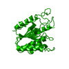
| ||||||||
| Unit cell |
|
-Components
| #1: Protein | Mass: 25986.453 Da / Num. of mol.: 1 / Mutation: T103S Source method: isolated from a genetically manipulated source Source: (gene. exp.)  References: UniProt: Q29460, 1-alkyl-2-acetylglycerophosphocholine esterase |
|---|---|
| #2: Water | ChemComp-HOH /  Mass: 18.015 Da / Num. of mol.: 133 / Source method: isolated from a natural source / Formula: H2O Mass: 18.015 Da / Num. of mol.: 133 / Source method: isolated from a natural source / Formula: H2O |
-Experimental details
-Experiment
| Experiment | Method: X-RAY DIFFRACTION / Number of used crystals: 2 |
|---|
- Sample preparation
| Crystal | Density Matthews: 2 Å3/Da / Density % sol: 39 % | ||||||||||||||||||||
|---|---|---|---|---|---|---|---|---|---|---|---|---|---|---|---|---|---|---|---|---|---|
| Crystal grow | pH: 6.8 / Details: pH 6.8 | ||||||||||||||||||||
| Crystal | *PLUS | ||||||||||||||||||||
| Crystal grow | *PLUS Method: vapor diffusion, sitting drop | ||||||||||||||||||||
| Components of the solutions | *PLUS
|
-Data collection
| Diffraction | Mean temperature: 100 K |
|---|---|
| Diffraction source | Wavelength: 1.5418 |
| Detector | Type: MARRESEARCH / Detector: IMAGE PLATE / Date: Nov 1, 1997 / Details: MSC MIRRORS |
| Radiation | Monochromator: GRAPHITE(002) / Monochromatic (M) / Laue (L): M / Scattering type: x-ray |
| Radiation wavelength | Wavelength: 1.5418 Å / Relative weight: 1 |
| Reflection | Resolution: 2.4→20 Å / Num. obs: 10257 / % possible obs: 92 % / Observed criterion σ(I): 0 / Redundancy: 10.7 % / Rsym value: 0.041 / Net I/σ(I): 18.3 |
| Reflection shell | Resolution: 2.3→2.38 Å / Rsym value: 0.161 / % possible all: 87.6 |
- Processing
| Software |
| ||||||||||||||||||||||||||||||||||||||||||||||||||||||||||||||||||||||||||||||||||||
|---|---|---|---|---|---|---|---|---|---|---|---|---|---|---|---|---|---|---|---|---|---|---|---|---|---|---|---|---|---|---|---|---|---|---|---|---|---|---|---|---|---|---|---|---|---|---|---|---|---|---|---|---|---|---|---|---|---|---|---|---|---|---|---|---|---|---|---|---|---|---|---|---|---|---|---|---|---|---|---|---|---|---|---|---|---|
| Refinement | Method to determine structure: MOLECULAR REPLACEMENT Starting model: PAF ACETYLHYDROLASE Resolution: 2.4→8 Å / σ(F): 0
| ||||||||||||||||||||||||||||||||||||||||||||||||||||||||||||||||||||||||||||||||||||
| Displacement parameters | Biso mean: 35.23 Å2 | ||||||||||||||||||||||||||||||||||||||||||||||||||||||||||||||||||||||||||||||||||||
| Refinement step | Cycle: LAST / Resolution: 2.4→8 Å
| ||||||||||||||||||||||||||||||||||||||||||||||||||||||||||||||||||||||||||||||||||||
| Refine LS restraints |
| ||||||||||||||||||||||||||||||||||||||||||||||||||||||||||||||||||||||||||||||||||||
| Software | *PLUS Name: REFMAC / Classification: refinement | ||||||||||||||||||||||||||||||||||||||||||||||||||||||||||||||||||||||||||||||||||||
| Refinement | *PLUS Rfactor obs: 0.202 | ||||||||||||||||||||||||||||||||||||||||||||||||||||||||||||||||||||||||||||||||||||
| Solvent computation | *PLUS | ||||||||||||||||||||||||||||||||||||||||||||||||||||||||||||||||||||||||||||||||||||
| Displacement parameters | *PLUS | ||||||||||||||||||||||||||||||||||||||||||||||||||||||||||||||||||||||||||||||||||||
| Refine LS restraints | *PLUS Type: p_planar_d / Dev ideal: 0.035 |
