 Movie
Movie Controller

[English] 日本語
 Yorodumi
- PDB-1es9: X-RAY CRYSTAL STRUCTURE OF R22K MUTANT OF THE MAMMALIAN BRAIN PLA... -
+ Open data

- Basic information
| Entry | Database: PDB / ID: 1es9 | ||||||
|---|---|---|---|---|---|---|---|
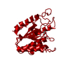
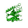
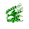
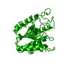
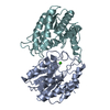
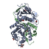
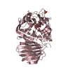
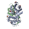
| Title | X-RAY CRYSTAL STRUCTURE OF R22K MUTANT OF THE MAMMALIAN BRAIN PLATELET-ACTIVATING FACTOR ACETYLHYDROLASES (PAF-AH) | ||||||
 Components Components | PLATELET-ACTIVATING FACTOR ACETYLHYDROLASE IB GAMMA SUBUNIT | ||||||
 Keywords Keywords | HYDROLASE / alpha/beta hydrolase fold | ||||||
| Function / homology |  Function and homology information Function and homology informationplatelet-activating factor acetyltransferase activity / 1-alkyl-2-acetylglycerophosphocholine esterase / 1-alkyl-2-acetylglycerophosphocholine esterase complex / COPI-independent Golgi-to-ER retrograde traffic / 1-alkyl-2-acetylglycerophosphocholine esterase activity / lipid catabolic process / spermatogenesis / protein heterodimerization activity / protein homodimerization activity / cytoplasm Similarity search - Function | ||||||
| Biological species |  | ||||||
| Method |  X-RAY DIFFRACTION / SYNCHROTRON / Resolution: 1.3 Å X-RAY DIFFRACTION / SYNCHROTRON / Resolution: 1.3 Å | ||||||
 Authors Authors | McMullen, T.W.P. / Li, J. / Sheffield, P.J. / Aoki, J. / Martin, T.W. / Arai, H. / Inoue, K. / Derewenda, Z.S. | ||||||
 Citation Citation | Journal: Protein Eng. / Year: 2000 Title: The functional implications of the dimerization of the catalytic subunits of the mammalian brain platelet-activating factor acetylhydrolase (Ib). Authors: McMullen, T.W. / Li, J. / Sheffield, P.J. / Aoki, J. / Martin, T.W. / Arai, H. / Inoue, K. / Derewenda, Z.S. #1: Journal: Nature / Year: 1997Title: Brain Acetylhydrolase that Inactivates Platelet-activating Factor is a G-protein-like trimer Authors: Ho, Y.S. / Swenson, L. / Derewenda, U. / Serre, L. / Wei, Y. / Dauter, Z. / Hattori, M. / Adachi, T. / Aoki, J. / Arai, H. / Inoue, K. / Derewenda, Z.S. | ||||||
| History |
|
- Structure visualization
| Structure viewer | Molecule: MolmilJmol/JSmol |
|---|
- Downloads & links
-Download
| PDBx/mmCIF format | 1es9.cif.gz | 62.5 KB | Display | PDBx/mmCIF format |
|---|---|---|---|---|
| PDB format | pdb1es9.ent.gz | 45.2 KB | Display | PDB format |
| PDBx/mmJSON format | 1es9.json.gz | Tree view | PDBx/mmJSON format | |
| Others |  Other downloads Other downloads |
-Validation report
| Arichive directory | https://data.pdbj.org/pub/pdb/validation_reports/es/1es9ftp://data.pdbj.org/pub/pdb/validation_reports/es/1es9 | HTTPS FTP |
|---|
-Related structure data
| Related structure data | |
|---|---|
| Similar structure data |


-Links
 PDBj
PDBj- Assembly
| Deposited unit | 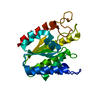
| ||||||||
|---|---|---|---|---|---|---|---|---|---|
| 1 | 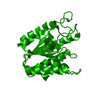
| ||||||||
| Unit cell |
| ||||||||
| Details | The biological assembly is a dimer |
-Components
| #1: Protein | Mass: 25875.350 Da / Num. of mol.: 1 / Mutation: R22K / Source method: isolated from a natural source / Source: (natural) References: UniProt: Q29460, 1-alkyl-2-acetylglycerophosphocholine esterase |
|---|---|
| #2: Water | ChemComp-HOH /  Mass: 18.015 Da / Num. of mol.: 297 / Source method: isolated from a natural source / Formula: H2O Mass: 18.015 Da / Num. of mol.: 297 / Source method: isolated from a natural source / Formula: H2O |
-Experimental details
-Experiment
| Experiment | Method: X-RAY DIFFRACTION / Number of used crystals: 1 |
|---|
- Sample preparation
| Crystal | Density Matthews: 2.66 Å3/Da / Density % sol: 53.77 % | ||||||||||||||||||||
|---|---|---|---|---|---|---|---|---|---|---|---|---|---|---|---|---|---|---|---|---|---|
| Crystal grow | Temperature: 293 K / Method: vapor diffusion, hanging drop / pH: 6.8 Details: 1.6M Ammonium sulphate, 100mM sodium acetate, pH 6.8, VAPOR DIFFUSION, HANGING DROP, temperature 20K | ||||||||||||||||||||
| Crystal grow | *PLUS Method: vapor diffusion, sitting drop / Details: Ho, Y.S., (1999) Protein Eng., 12, 693. | ||||||||||||||||||||
| Components of the solutions | *PLUS
|
-Data collection
| Diffraction | Mean temperature: 100 K |
|---|---|
| Diffraction source | Source: SYNCHROTRON / Site: NSLS  / Beamline: X9B / Wavelength: 0.9724 / Beamline: X9B / Wavelength: 0.9724 |
| Detector | Type: MARRESEARCH / Detector: IMAGE PLATE / Date: Jan 14, 1999 |
| Radiation | Protocol: SINGLE WAVELENGTH / Monochromatic (M) / Laue (L): M / Scattering type: x-ray |
| Radiation wavelength | Wavelength: 0.9724 Å / Relative weight: 1 |
| Reflection | Resolution: 1.2→20 Å / Num. all: 85358 / Num. obs: 72050 / % possible obs: 84.4 % / Observed criterion σ(F): 1 / Observed criterion σ(I): 1 / Redundancy: 5.4 % / Biso Wilson estimate: 12.2 Å2 / Rmerge(I) obs: 0.069 / Net I/σ(I): 32 |
| Reflection shell | Resolution: 1.2→1.24 Å / Redundancy: 2 % / Rmerge(I) obs: 0.448 / Num. unique all: 7738 / % possible all: 87.4 |
| Reflection | *PLUS Num. obs: 85358 / % possible obs: 98.7 % / Num. measured all: 461423 |
| Reflection shell | *PLUS % possible obs: 90.4 % / Rmerge(I) obs: 0.366 |
- Processing
| Software |
| |||||||||||||||||||||||||
|---|---|---|---|---|---|---|---|---|---|---|---|---|---|---|---|---|---|---|---|---|---|---|---|---|---|---|
| Refinement | Resolution: 1.3→10 Å / σ(F): 2 / σ(I): 2 / Stereochemistry target values: Engh & Huber
| |||||||||||||||||||||||||
| Refinement step | Cycle: LAST / Resolution: 1.3→10 Å
| |||||||||||||||||||||||||
| Refine LS restraints |
| |||||||||||||||||||||||||
| Software | *PLUS Name: REFMAC / Classification: refinement | |||||||||||||||||||||||||
| Refine LS restraints | *PLUS Type: p_planar_d / Dev ideal: 0.024 |
