 Movie
Movie Controller

[English] 日本語
 Yorodumi
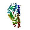
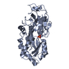
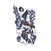
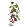
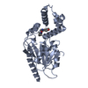
- PDB-1bo4: CRYSTAL STRUCTURE OF A GCN5-RELATED N-ACETYLTRANSFERASE: SERRATIA... -
+ Open data

- Basic information
| Entry | Database: PDB / ID: 1bo4 | ||||||
|---|---|---|---|---|---|---|---|
| Title | CRYSTAL STRUCTURE OF A GCN5-RELATED N-ACETYLTRANSFERASE: SERRATIA MARESCENS AMINOGLYCOSIDE 3-N-ACETYLTRANSFERASE | ||||||
 Components Components | PROTEIN (SERRATIA MARCESCENS AMINOGLYCOSIDE-3-N-ACETYLTRANSFERASE) | ||||||
 Keywords Keywords | TRANSFERASE / AMINOGLYCOSIDE 3-N-ACETYLTRANSFERASE / EUBACTERIAL AMINOGLYCOSIDE RESISTANCE / GCN5-RELATED N-ACETYLTRANSFERASE / COA-BINDING | ||||||
| Function / homology |  Function and homology information Function and homology informationacyltransferase activity, transferring groups other than amino-acyl groups Similarity search - Function | ||||||
| Biological species |  Serratia marcescens (bacteria) Serratia marcescens (bacteria) | ||||||
| Method |  X-RAY DIFFRACTION / SYNCHROTRON / MAD / Resolution: 2.3 Å X-RAY DIFFRACTION / SYNCHROTRON / MAD / Resolution: 2.3 Å | ||||||
 Authors Authors | Wolf, E. / Vassilev, A. / Makino, Y. / Sali, A. / Nakatani, Y. / Burley, S.K. | ||||||
 Citation Citation | Journal: Cell(Cambridge,Mass.) / Year: 1998 Title: Crystal structure of a GCN5-related N-acetyltransferase: Serratia marcescens aminoglycoside 3-N-acetyltransferase. Authors: Wolf, E. / Vassilev, A. / Makino, Y. / Sali, A. / Nakatani, Y. / Burley, S.K. | ||||||
| History |
|
- Structure visualization
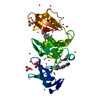
| Structure viewer | Molecule: MolmilJmol/JSmol |
|---|
- Downloads & links
-Download
| PDBx/mmCIF format | 1bo4.cif.gz | 74.5 KB | Display | PDBx/mmCIF format |
|---|---|---|---|---|
| PDB format | pdb1bo4.ent.gz | 55.7 KB | Display | PDB format |
| PDBx/mmJSON format | 1bo4.json.gz | Tree view | PDBx/mmJSON format | |
| Others |  Other downloads Other downloads |
-Validation report
| Arichive directory | https://data.pdbj.org/pub/pdb/validation_reports/bo/1bo4ftp://data.pdbj.org/pub/pdb/validation_reports/bo/1bo4 | HTTPS FTP |
|---|
-Related structure data
| Similar structure data |
|---|


-Links
 PDBj
PDBj


- Assembly
| Deposited unit | 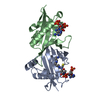
| ||||||||
|---|---|---|---|---|---|---|---|---|---|
| 1 |
| ||||||||
| Unit cell |
| ||||||||
| Noncrystallographic symmetry (NCS) | NCS oper: (Code: given Matrix: (-0.999954, 9.4E-5, 0.009258), Vector: |
-Components
| #1: Protein | Mass: 18487.984 Da / Num. of mol.: 2 Source method: isolated from a genetically manipulated source Source: (gene. exp.) Serratia marcescens (bacteria) / Cell: EUBACTERIA / Cellular location: CYTOPLASM / Gene: AACC1 / Species (production host): Escherichia coli / Cellular location (production host): CYTOPLASM / Production host: #2: Chemical | ChemComp-SPD / |   Mass: 145.246 Da / Num. of mol.: 1 / Source method: obtained synthetically / Formula: C7H19N3 Mass: 145.246 Da / Num. of mol.: 1 / Source method: obtained synthetically / Formula: C7H19N3#3: Chemical |   Mass: 767.534 Da / Num. of mol.: 2 / Source method: obtained synthetically / Formula: C21H36N7O16P3S Mass: 767.534 Da / Num. of mol.: 2 / Source method: obtained synthetically / Formula: C21H36N7O16P3S#4: Water | ChemComp-HOH / |  Mass: 18.015 Da / Num. of mol.: 253 / Source method: isolated from a natural source / Formula: H2O Mass: 18.015 Da / Num. of mol.: 253 / Source method: isolated from a natural source / Formula: H2O |
|---|
-Experimental details
-Experiment
| Experiment | Method: X-RAY DIFFRACTION / Number of used crystals: 1 |
|---|
- Sample preparation
| Crystal | Density Matthews: 2.7 Å3/Da / Density % sol: 54 % | ||||||||||||||||||||||||||||||||||||||||||||||||||||||||||||||||||||||||||||||||||||
|---|---|---|---|---|---|---|---|---|---|---|---|---|---|---|---|---|---|---|---|---|---|---|---|---|---|---|---|---|---|---|---|---|---|---|---|---|---|---|---|---|---|---|---|---|---|---|---|---|---|---|---|---|---|---|---|---|---|---|---|---|---|---|---|---|---|---|---|---|---|---|---|---|---|---|---|---|---|---|---|---|---|---|---|---|---|
| Crystal grow | Temperature: 277 K / Method: vapor diffusion, sitting drop / pH: 7.8 Details: SITTING DROP, 4 DEGREES C, 3.5MG/ML PROTEIN + 5MM COA, 100MM TRIS-HCL PH7.8, 0.8% PEG4K, 18% T-BUTANOL, 20MM SRCL2, 7% DIOXANE, 5% 2,4-METHYLPENTANEDIOL, 40MM HEXANEDIOL, 10MM ...Details: SITTING DROP, 4 DEGREES C, 3.5MG/ML PROTEIN + 5MM COA, 100MM TRIS-HCL PH7.8, 0.8% PEG4K, 18% T-BUTANOL, 20MM SRCL2, 7% DIOXANE, 5% 2,4-METHYLPENTANEDIOL, 40MM HEXANEDIOL, 10MM DITHIOTHREITOL, 2MM TRIS(2-CARBOXY- ETHYL)PHOSPHINE-HCL, 10MM SPERMIDINE, 2.5% GLYCEROL, vapor diffusion - sitting drop, temperature 277K | ||||||||||||||||||||||||||||||||||||||||||||||||||||||||||||||||||||||||||||||||||||
| Crystal grow | *PLUS Temperature: 4 ℃ | ||||||||||||||||||||||||||||||||||||||||||||||||||||||||||||||||||||||||||||||||||||
| Components of the solutions | *PLUS
|
-Data collection
| Diffraction | Mean temperature: 100 K | ||||||||||||
|---|---|---|---|---|---|---|---|---|---|---|---|---|---|
| Diffraction source | Source: SYNCHROTRON / Site: CHESS  / Beamline: F2 / Wavelength: 0.9793, 0.9703, 0.9668 / Beamline: F2 / Wavelength: 0.9793, 0.9703, 0.9668 | ||||||||||||
| Detector | Detector: CCD / Date: May 15, 1998 | ||||||||||||
| Radiation | Protocol: MAD / Monochromatic (M) / Laue (L): M / Scattering type: x-ray | ||||||||||||
| Radiation wavelength |
| ||||||||||||
| Reflection | Resolution: 2.3→20 Å / Num. obs: 16291 / % possible obs: 92.9 % / Observed criterion σ(I): 2 / Redundancy: 7.3 % / Biso Wilson estimate: 27 Å2 / Rsym value: 0.052 / Net I/σ(I): 7.6 | ||||||||||||
| Reflection shell | Resolution: 2.3→2.42 Å / Redundancy: 7.2 % / Mean I/σ(I) obs: 6.4 / Rsym value: 0.093 / % possible all: 92.9 | ||||||||||||
| Reflection | *PLUS Num. measured all: 118408 / Rmerge(I) obs: 0.052 | ||||||||||||
| Reflection shell | *PLUS % possible obs: 92.9 % / Rmerge(I) obs: 0.093 |
- Processing
| Software |
| ||||||||||||||||||||||||||||||||||||||||||||||||||||||||||||||||||||||||||||||||
|---|---|---|---|---|---|---|---|---|---|---|---|---|---|---|---|---|---|---|---|---|---|---|---|---|---|---|---|---|---|---|---|---|---|---|---|---|---|---|---|---|---|---|---|---|---|---|---|---|---|---|---|---|---|---|---|---|---|---|---|---|---|---|---|---|---|---|---|---|---|---|---|---|---|---|---|---|---|---|---|---|---|
| Refinement | Method to determine structure: MAD / Resolution: 2.3→15 Å / Data cutoff high rms absF: 10000 / Isotropic thermal model: RESTRAINED / Cross valid method: THROUGHOUT / σ(F): 2
| ||||||||||||||||||||||||||||||||||||||||||||||||||||||||||||||||||||||||||||||||
| Solvent computation | Bsol: 49.7 Å2 / ksol: 0.37 e/Å3 | ||||||||||||||||||||||||||||||||||||||||||||||||||||||||||||||||||||||||||||||||
| Displacement parameters | Biso mean: 24.9 Å2
| ||||||||||||||||||||||||||||||||||||||||||||||||||||||||||||||||||||||||||||||||
| Refinement step | Cycle: LAST / Resolution: 2.3→15 Å
| ||||||||||||||||||||||||||||||||||||||||||||||||||||||||||||||||||||||||||||||||
| Refine LS restraints |
| ||||||||||||||||||||||||||||||||||||||||||||||||||||||||||||||||||||||||||||||||
| Refine LS restraints NCS | NCS model details: RESTRAINTS | ||||||||||||||||||||||||||||||||||||||||||||||||||||||||||||||||||||||||||||||||
| LS refinement shell | Resolution: 2.3→2.33 Å / Total num. of bins used: 31
| ||||||||||||||||||||||||||||||||||||||||||||||||||||||||||||||||||||||||||||||||
| Xplor file |
| ||||||||||||||||||||||||||||||||||||||||||||||||||||||||||||||||||||||||||||||||
| Software | *PLUS Name: CNS / Version: 0.3C / Classification: refinement | ||||||||||||||||||||||||||||||||||||||||||||||||||||||||||||||||||||||||||||||||
| Refine LS restraints | *PLUS
|
