 Movie
Movie Controller


+ Open data

- Basic information
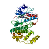
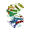
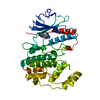
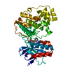
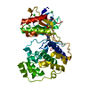
| Entry | Database: PDB / ID: 1bmk | ||||||
|---|---|---|---|---|---|---|---|
| Title | THE COMPLEX STRUCTURE OF THE MAP KINASE P38/SB218655 | ||||||
 Components Components | PROTEIN (MAP KINASE P38) | ||||||
 Keywords Keywords |  TRANSFERASE / INHIBITORS / MAP KINASE / SERINE/ THREONINE-PROTEIN KINASE / P38 TRANSFERASE / INHIBITORS / MAP KINASE / SERINE/ THREONINE-PROTEIN KINASE / P38 | ||||||
| Function / homology |  Function and homology information Function and homology informationpositive regulation of cyclase activity / Activation of PPARGC1A (PGC-1alpha) by phosphorylation / CD163 mediating an anti-inflammatory response / regulation of synaptic membrane adhesion / stress-induced premature senescence / cell surface receptor protein serine/threonine kinase signaling pathway / 3'-UTR-mediated mRNA stabilization / KSRP (KHSRP) binds and destabilizes mRNA / DSCAM interactions / cartilage condensation ...positive regulation of cyclase activity / Activation of PPARGC1A (PGC-1alpha) by phosphorylation / CD163 mediating an anti-inflammatory response / regulation of synaptic membrane adhesion / stress-induced premature senescence / cell surface receptor protein serine/threonine kinase signaling pathway / 3'-UTR-mediated mRNA stabilization / KSRP (KHSRP) binds and destabilizes mRNA / DSCAM interactions / cartilage condensation / cellular response to UV-B / Platelet sensitization by LDL / stress-activated protein kinase signaling cascade / mitogen-activated protein kinase p38 binding / positive regulation of myoblast fusion / positive regulation of muscle cell differentiation / negative regulation of hippo signaling / positive regulation of myotube differentiation / NFAT protein binding / Myogenesis / glucose import / response to dietary excess / Activation of the AP-1 family of transcription factors / ERK/MAPK targets / regulation of cytokine production involved in inflammatory response / p38MAPK cascade / fatty acid oxidation / cellular response to lipoteichoic acid / MAP kinase kinase activity / response to muramyl dipeptide / RHO GTPases Activate NADPH Oxidases / regulation of ossification / MAP kinase activity / mitogen-activated protein kinase / cellular response to vascular endothelial growth factor stimulus / positive regulation of myoblast differentiation / chondrocyte differentiation / vascular endothelial growth factor receptor signaling pathway / stress-activated MAPK cascade / skeletal muscle tissue development / signal transduction in response to DNA damage / lipopolysaccharide-mediated signaling pathway / negative regulation of inflammatory response to antigenic stimulus / positive regulation of cardiac muscle cell proliferation / p38MAPK events / striated muscle cell differentiation / response to muscle stretch / positive regulation of brown fat cell differentiation / positive regulation of interleukin-12 production / osteoclast differentiation / positive regulation of erythrocyte differentiation / placenta development / DNA damage checkpoint signaling / activated TAK1 mediates p38 MAPK activation / cellular response to ionizing radiation / stem cell differentiation / positive regulation of glucose import / negative regulation of canonical Wnt signaling pathway / response to insulin / NOD1/2 Signaling Pathway / bone development / cell morphogenesis / platelet activation / osteoblast differentiation / cellular response to virus / VEGFA-VEGFR2 Pathway / spindle pole / positive regulation of protein import into nucleus / ADP signalling through P2Y purinoceptor 1 / glucose metabolic process / positive regulation of reactive oxygen species metabolic process / chemotaxis / cellular senescence / cellular response to tumor necrosis factor / protein phosphatase binding / peptidyl-serine phosphorylation / angiogenesis / Oxidative Stress Induced Senescence / secretory granule lumen / cellular response to lipopolysaccharide / Regulation of TP53 Activity through Phosphorylation / ficolin-1-rich granule lumen / transcription by RNA polymerase II / cell surface receptor signaling pathway / intracellular signal transduction / nuclear speck / protein serine kinase activity / protein serine/threonine kinase activity / glutamatergic synapse / apoptotic process / Neutrophil degranulation / positive regulation of gene expression / regulation of transcription by RNA polymerase II / enzyme binding / signal transduction / positive regulation of transcription by RNA polymerase II / mitochondrion / extracellular region / nucleoplasm / ATP bindingSimilarity search - Function | ||||||
| Biological species |  Homo sapiens (human) Homo sapiens (human) | ||||||
| Method | X-RAY DIFFRACTION / MOLECULAR REPLACEMENT / Resolution: 2.4 Å | ||||||
 Authors Authors | Wang, Z. / Canagarajah, B. / Boehm, J.C. / Kassis, S. / Cobb, M.H. / Young, P.R. / Abdel-Meguid, S. / Adams, J.L. / Goldsmith, E.J. | ||||||
 Citation Citation | Journal: Structure / Year: 1998 Title: Structural basis of inhibitor selectivity in MAP kinases. Authors: Wang, Z. / Canagarajah, B.J. / Boehm, J.C. / Kassisa, S. / Cobb, M.H. / Young, P.R. / Abdel-Meguid, S. / Adams, J.L. / Goldsmith, E.J. #1: Journal: J.Biol.Chem. / Year: 1995Title: Pro-Inflammatory Cytokines and Environmental Stress Cause P38 Mitogen- Activated Protein Kinase Activation by Dual Phosphorylation on Tyrosine and Threonine Authors: Raingeaud, J. / Gupta, S. / Rogers, J.S. / Dickens, M. / Han, J. / Ulevitch, R.J. / Davis, R.J. #2: Journal: Science / Year: 1994Title: A Map Kinase Targeted by Endotoxin and Hyperosmolarity in Mammalian Cells Authors: Han, J. / Lee, J.D. / Bibbs, L. / Ulevitch, R.J. #3: Journal: Nature / Year: 1994Title: A Protein Kinase Involved in the Regulation of Inflammatory Cytokine Biosynthesis Authors: Lee, J.C. / Laydon, J.T. / Mcdonnell, P.C. / Gallagher, T.F. / Kumar, S. / Green, D. / Mcnulty, D. / Blumenthal, M.J. / Heys, J.R. / Landvatter, S.W. / Strickler, J.E. / Mclaughlin, M.M. / ...Authors: Lee, J.C. / Laydon, J.T. / Mcdonnell, P.C. / Gallagher, T.F. / Kumar, S. / Green, D. / Mcnulty, D. / Blumenthal, M.J. / Heys, J.R. / Landvatter, S.W. / Strickler, J.E. / Mclaughlin, M.M. / Siemens, I.R. / Fisher, S.M. / Livi, G.P. / White, J.R. / Adams, J.L. / Young, P.R. | ||||||
| History |
|
- Structure visualization
| Structure viewer | Molecule: MolmilJmol/JSmol |
|---|
- Downloads & links
-Download
| PDBx/mmCIF format | 1bmk.cif.gz | 86.3 KB | Display | PDBx/mmCIF format |
|---|---|---|---|---|
| PDB format | pdb1bmk.ent.gz | 64.6 KB | Display | PDB format |
| PDBx/mmJSON format | 1bmk.json.gz | Tree view | PDBx/mmJSON format | |
| Others |  Other downloads Other downloads |
-Validation report
| Arichive directory | https://data.pdbj.org/pub/pdb/validation_reports/bm/1bmkftp://data.pdbj.org/pub/pdb/validation_reports/bm/1bmk | HTTPS FTP |
|---|
-Related structure data


-Links
 PDBj
PDBj


- Assembly
| Deposited unit | 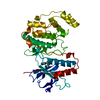
| ||||||||
|---|---|---|---|---|---|---|---|---|---|
| 1 |
| ||||||||
| Unit cell |
|
-Components
| #1: Protein | Mass: 43378.312 Da / Num. of mol.: 1 / Mutation: 19 RESIDUES INSERTED AT N-TERMINUS Source method: isolated from a genetically manipulated source Details: SB218655 PYRIDINYLIMIDAZOLE / Source: (gene. exp.) Homo sapiens (human) / Strain: BL21 (DE3) / Description: YES / Plasmid: PET15B / Species (production host): Escherichia coli / Cell line (production host): BL21(DE3) / Production host:  Escherichia coli BL21(DE3) (bacteria) / Strain (production host): BL21 (DE3) / References: UniProt: Q16539 Escherichia coli BL21(DE3) (bacteria) / Strain (production host): BL21 (DE3) / References: UniProt: Q16539 |
|---|---|
| #2: Chemical | ChemComp-SB5 /   Mass: 309.341 Da / Num. of mol.: 1 / Source method: obtained synthetically / Formula: C17H16FN5 Mass: 309.341 Da / Num. of mol.: 1 / Source method: obtained synthetically / Formula: C17H16FN5 |
| #3: Water | ChemComp-HOH / Water Mass: 18.015 Da / Num. of mol.: 94 / Source method: isolated from a natural source / Formula: H2O Mass: 18.015 Da / Num. of mol.: 94 / Source method: isolated from a natural source / Formula: H2O |
-Experimental details
-Experiment
| Experiment | Method: X-RAY DIFFRACTION / Number of used crystals: 1 |
|---|
- Sample preparation
| Crystal | Density Matthews: 2.92 Å3/Da / Density % sol: 58 % | ||||||||||||||||||||||||||||||||||||||||||||||||||||||||||||||||||||||||
|---|---|---|---|---|---|---|---|---|---|---|---|---|---|---|---|---|---|---|---|---|---|---|---|---|---|---|---|---|---|---|---|---|---|---|---|---|---|---|---|---|---|---|---|---|---|---|---|---|---|---|---|---|---|---|---|---|---|---|---|---|---|---|---|---|---|---|---|---|---|---|---|---|---|
| Crystal grow | pH: 7.4 Details: 18% PEG 8000, 0.2M MG(OAC)2, 0.1M HEPES, PH7.0, pH 7.4 | ||||||||||||||||||||||||||||||||||||||||||||||||||||||||||||||||||||||||
| Crystal grow | *PLUS Method: vapor diffusionDetails: Wang, Z., (1997) Proc. Natl. Acad. Sci. USA, 94, 2327. | ||||||||||||||||||||||||||||||||||||||||||||||||||||||||||||||||||||||||
| Components of the solutions | *PLUS
|
-Data collection
| Diffraction | Mean temperature: 100 K |
|---|---|
| Diffraction source | Source: ROTATING ANODE / Type: RIGAKU RU300 / Wavelength: 1.54 |
| Detector | Type: RIGAKU RAXIS IIC / Detector: IMAGE PLATE / Date: Mar 3, 1997 / Details: MIRRORS |
| Radiation | Protocol: SINGLE WAVELENGTH / Monochromatic (M) / Laue (L): M / Scattering type: x-ray |
| Radiation wavelength | Wavelength: 1.54 Å / Relative weight: 1 |
| Reflection | Resolution: 2.4→35.1 Å / Num. obs: 19500 / % possible obs: 99.4 % / Observed criterion σ(I): -3 / Redundancy: 10.3 % / Rsym value: 0.05 / Net I/σ(I): 22.6 |
| Reflection shell | Resolution: 2.4→2.49 Å / Mean I/σ(I) obs: 6.9 / Rsym value: 0.201 / % possible all: 96 |
| Reflection | *PLUS Num. measured all: 200387 / Rmerge(I) obs: 0.05 |
- Processing
| Software |
| ||||||||||||||||||||||||||||||||||||||||||||||||||||||||||||
|---|---|---|---|---|---|---|---|---|---|---|---|---|---|---|---|---|---|---|---|---|---|---|---|---|---|---|---|---|---|---|---|---|---|---|---|---|---|---|---|---|---|---|---|---|---|---|---|---|---|---|---|---|---|---|---|---|---|---|---|---|---|
| Refinement | Method to determine structure: MOLECULAR REPLACEMENT Starting model: MAP KINASE P38 Resolution: 2.4→20 Å / σ(F): 2
| ||||||||||||||||||||||||||||||||||||||||||||||||||||||||||||
| Displacement parameters | Biso mean: 41.6 Å2 | ||||||||||||||||||||||||||||||||||||||||||||||||||||||||||||
| Refinement step | Cycle: LAST / Resolution: 2.4→20 Å
| ||||||||||||||||||||||||||||||||||||||||||||||||||||||||||||
| Refine LS restraints |
| ||||||||||||||||||||||||||||||||||||||||||||||||||||||||||||
| Xplor file |
| ||||||||||||||||||||||||||||||||||||||||||||||||||||||||||||
| Software | *PLUS Name: X-PLOR / Classification: refinement | ||||||||||||||||||||||||||||||||||||||||||||||||||||||||||||
| Refinement | *PLUS Highest resolution: 2.4 Å / σ(F): 2 / % reflection Rfree: 10 % | ||||||||||||||||||||||||||||||||||||||||||||||||||||||||||||
| Solvent computation | *PLUS | ||||||||||||||||||||||||||||||||||||||||||||||||||||||||||||
| Displacement parameters | *PLUS Biso mean: 41.6 Å2 | ||||||||||||||||||||||||||||||||||||||||||||||||||||||||||||
| Refine LS restraints | *PLUS
|
