 Movie
Movie Controller

[English] 日本語
 Yorodumi
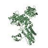
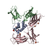
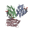
- PDB-1bkn: CRYSTAL STRUCTURE OF AN N-TERMINAL 40KD FRAGMENT OF E. COLI DNA M... -
+ Open data

- Basic information
| Entry | Database: PDB / ID: 1bkn | ||||||
|---|---|---|---|---|---|---|---|
| Title | CRYSTAL STRUCTURE OF AN N-TERMINAL 40KD FRAGMENT OF E. COLI DNA MISMATCH REPAIR PROTEIN MUTL | ||||||
 Components Components | MUTL | ||||||
 Keywords Keywords | DNA REPAIR / ATPASE / DNA BINDING | ||||||
| Function / homology |  Function and homology information Function and homology informationsingle-stranded DNA-dependent ATP-dependent DNA helicase complex / mismatch repair involved in maintenance of fidelity involved in DNA-dependent DNA replication / mismatch repair complex / regulation of DNA recombination / mismatched DNA binding / ATP-dependent DNA damage sensor activity / mismatch repair / ATP hydrolysis activity / DNA binding / ATP binding / identical protein binding Similarity search - Function | ||||||
| Biological species |  | ||||||
| Method |  X-RAY DIFFRACTION / SIRAS / Resolution: 2.9 Å X-RAY DIFFRACTION / SIRAS / Resolution: 2.9 Å | ||||||
 Authors Authors | Yang, W. / Ban, C. | ||||||
 Citation Citation | Journal: Cell(Cambridge,Mass.) / Year: 1998 Title: Crystal structure and ATPase activity of MutL: implications for DNA repair and mutagenesis. Authors: Ban, C. / Yang, W. | ||||||
| History |
|
- Structure visualization
| Structure viewer | Molecule: MolmilJmol/JSmol |
|---|
- Downloads & links
-Download
| PDBx/mmCIF format | 1bkn.cif.gz | 117.5 KB | Display | PDBx/mmCIF format |
|---|---|---|---|---|
| PDB format | pdb1bkn.ent.gz | 90.6 KB | Display | PDB format |
| PDBx/mmJSON format | 1bkn.json.gz | Tree view | PDBx/mmJSON format | |
| Others |  Other downloads Other downloads |
-Validation report
| Arichive directory | https://data.pdbj.org/pub/pdb/validation_reports/bk/1bknftp://data.pdbj.org/pub/pdb/validation_reports/bk/1bkn | HTTPS FTP |
|---|
-Related structure data
| Similar structure data |
|---|


-Links
 PDBj
PDBj- Assembly
| Deposited unit | 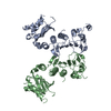
| ||||||||
|---|---|---|---|---|---|---|---|---|---|
| 1 |
| ||||||||
| Unit cell |
| ||||||||
| Noncrystallographic symmetry (NCS) | NCS oper: (Code: given Matrix: (0.041784, 0.989309, -0.139721), Vector: |
-Components
| #1: Protein | Mass: 39299.602 Da / Num. of mol.: 2 / Fragment: N-TERMINAL 40KD FRAGMENT Source method: isolated from a genetically manipulated source Source: (gene. exp.) #2: Water | ChemComp-HOH / |  Mass: 18.015 Da / Num. of mol.: 55 / Source method: isolated from a natural source / Formula: H2O Mass: 18.015 Da / Num. of mol.: 55 / Source method: isolated from a natural source / Formula: H2OSequence details | THE FIRST THREE RESIDUES IN THE SEQUENCE ARE NOT ENCODED BY E. COLI MUTL GENE. THEY ARE FUSED INTO ...THE FIRST THREE RESIDUES IN THE SEQUENCE ARE NOT ENCODED BY E. COLI MUTL GENE. THEY ARE FUSED INTO MUTL PROTEIN DUE TO CLONING AND EXPRESSION | |
|---|
-Experimental details
-Experiment
| Experiment | Method: X-RAY DIFFRACTION / Number of used crystals: 1 |
|---|
- Sample preparation
| Crystal | Density Matthews: 2.86 Å3/Da / Density % sol: 57 % Description: DATA TO 2.9A ARE USED FOR THE STRUCTURE REFINEMENT. THE LAST SHELL (2.9A TO 3.0A) HAS RSYM OF 0.50, COMPLETENESS OF 98.3% AND I/SIGI OF 2.1. | ||||||||||||||||||||||||||||||
|---|---|---|---|---|---|---|---|---|---|---|---|---|---|---|---|---|---|---|---|---|---|---|---|---|---|---|---|---|---|---|---|
| Crystal grow | pH: 6.6 / Details: pH 6.6 | ||||||||||||||||||||||||||||||
| Crystal | *PLUS | ||||||||||||||||||||||||||||||
| Crystal grow | *PLUS Temperature: 20 ℃ / Method: vapor diffusion, hanging drop | ||||||||||||||||||||||||||||||
| Components of the solutions | *PLUS
|
-Data collection
| Diffraction | Mean temperature: 95 K |
|---|---|
| Diffraction source | Source: ROTATING ANODE / Type: RIGAKU RUH2R / Wavelength: 1.5418 |
| Detector | Type: RIGAKU RAXIS II / Detector: IMAGE PLATE / Date: Aug 1, 1997 / Details: MIRROR |
| Radiation | Monochromator: NI FILTER / Monochromatic (M) / Laue (L): M / Scattering type: x-ray |
| Radiation wavelength | Wavelength: 1.5418 Å / Relative weight: 1 |
| Reflection | Resolution: 2.7→20 Å / Num. obs: 23519 / % possible obs: 96.5 % / Observed criterion σ(I): 0 / Redundancy: 4.5 % / Biso Wilson estimate: 84.7 Å2 / Rsym value: 0.074 / Net I/σ(I): 8.3 |
| Reflection shell | Resolution: 2.7→2.8 Å / Redundancy: 2.7 % / Mean I/σ(I) obs: 1.1 / Rsym value: 0.7 / % possible all: 84.6 |
| Reflection | *PLUS Rmerge(I) obs: 0.074 |
| Reflection shell | *PLUS % possible obs: 84.6 % / Rmerge(I) obs: 0.696 |
- Processing
| Software |
| ||||||||||||||||||||||||||||||||||||||||||||||||||||||||||||||||||||||||||||||||
|---|---|---|---|---|---|---|---|---|---|---|---|---|---|---|---|---|---|---|---|---|---|---|---|---|---|---|---|---|---|---|---|---|---|---|---|---|---|---|---|---|---|---|---|---|---|---|---|---|---|---|---|---|---|---|---|---|---|---|---|---|---|---|---|---|---|---|---|---|---|---|---|---|---|---|---|---|---|---|---|---|---|
| Refinement | Method to determine structure: SIRAS / Resolution: 2.9→20 Å / Rfactor Rfree error: 0.01 / Data cutoff high absF: 10000000 / Data cutoff low absF: 0.001 / Isotropic thermal model: RESTRAINED / Cross valid method: THROUGHOUT / σ(F): 0
| ||||||||||||||||||||||||||||||||||||||||||||||||||||||||||||||||||||||||||||||||
| Displacement parameters | Biso mean: 73.2 Å2
| ||||||||||||||||||||||||||||||||||||||||||||||||||||||||||||||||||||||||||||||||
| Refine analyze |
| ||||||||||||||||||||||||||||||||||||||||||||||||||||||||||||||||||||||||||||||||
| Refinement step | Cycle: LAST / Resolution: 2.9→20 Å
| ||||||||||||||||||||||||||||||||||||||||||||||||||||||||||||||||||||||||||||||||
| Refine LS restraints |
| ||||||||||||||||||||||||||||||||||||||||||||||||||||||||||||||||||||||||||||||||
| LS refinement shell | Resolution: 2.9→3.08 Å / Rfactor Rfree error: 0.032 / Total num. of bins used: 6
| ||||||||||||||||||||||||||||||||||||||||||||||||||||||||||||||||||||||||||||||||
| Xplor file |
| ||||||||||||||||||||||||||||||||||||||||||||||||||||||||||||||||||||||||||||||||
| Software | *PLUS Name: X-PLOR / Version: 3.851 / Classification: refinement | ||||||||||||||||||||||||||||||||||||||||||||||||||||||||||||||||||||||||||||||||
| Refinement | *PLUS Rfactor Rfree: 0.3 | ||||||||||||||||||||||||||||||||||||||||||||||||||||||||||||||||||||||||||||||||
| Solvent computation | *PLUS | ||||||||||||||||||||||||||||||||||||||||||||||||||||||||||||||||||||||||||||||||
| Displacement parameters | *PLUS | ||||||||||||||||||||||||||||||||||||||||||||||||||||||||||||||||||||||||||||||||
| Refine LS restraints | *PLUS
|
