 Movie
Movie Controller


+ Open data

- Basic information
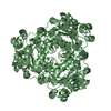
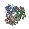
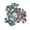
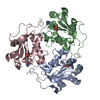
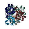
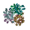
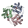
| Entry | Database: PDB / ID: 1be4 | ||||||
|---|---|---|---|---|---|---|---|
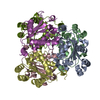
| Title | NUCLEOSIDE DIPHOSPHATE KINASE ISOFORM B FROM BOVINE RETINA | ||||||
 Components Components | NUCLEOSIDE DIPHOSPHATE TRANSFERASE | ||||||
 Keywords Keywords |  PHOSPHOTRANSFERASE PHOSPHOTRANSFERASE | ||||||
| Function / homology |  Function and homology information Function and homology informationDNA nuclease activity / nucleoside-diphosphate kinase / UTP biosynthetic process / CTP biosynthetic process / GTP biosynthetic process / nucleoside diphosphate kinase activity / ribosomal small subunit binding / positive regulation of DNA binding / lactation / positive regulation of epithelial cell proliferation ...DNA nuclease activity / nucleoside-diphosphate kinase / UTP biosynthetic process / CTP biosynthetic process / GTP biosynthetic process / nucleoside diphosphate kinase activity / ribosomal small subunit binding / positive regulation of DNA binding / lactation / positive regulation of epithelial cell proliferation / ruffle membrane / endocytosis / nervous system development / cell differentiation / GTP binding / magnesium ion binding / ATP binding / identical protein binding / nucleus / cytosolSimilarity search - Function | ||||||
| Biological species |  Bos taurus (cattle) Bos taurus (cattle) | ||||||
| Method | X-RAY DIFFRACTION / MOLECULAR REPLACEMENT / Resolution: 2.4 Å | ||||||
 Authors Authors | Ladner, J.E. / Abdulaev, N.G. / Kakuev, D.L. / Karaschuk, G.N. / Tordova, M. / Eisenstein, E. / Fujiwara, J.H. / Ridge, K.D. / Gilliland, G.L. | ||||||
 Citation Citation | Journal: Biochemistry / Year: 1998 Title: Nucleoside diphosphate kinase from bovine retina: purification, subcellular localization, molecular cloning, and three-dimensional structure. Authors: Abdulaev, N.G. / Karaschuk, G.N. / Ladner, J.E. / Kakuev, D.L. / Yakhyaev, A.V. / Tordova, M. / Gaidarov, I.O. / Popov, V.I. / Fujiwara, J.H. / Chinchilla, D. / Eisenstein, E. / Gilliland, G.L. / Ridge, K.D. | ||||||
| History |
|
- Structure visualization
| Structure viewer | Molecule: MolmilJmol/JSmol |
|---|
- Downloads & links
-Download
| PDBx/mmCIF format | 1be4.cif.gz | 111.8 KB | Display | PDBx/mmCIF format |
|---|---|---|---|---|
| PDB format | pdb1be4.ent.gz | 86.6 KB | Display | PDB format |
| PDBx/mmJSON format | 1be4.json.gz | Tree view | PDBx/mmJSON format | |
| Others |  Other downloads Other downloads |
-Validation report
| Arichive directory | https://data.pdbj.org/pub/pdb/validation_reports/be/1be4ftp://data.pdbj.org/pub/pdb/validation_reports/be/1be4 | HTTPS FTP |
|---|
-Related structure data
| Related structure data | 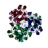 1nskS S: Starting model for refinement |
|---|---|
| Similar structure data |


-Links
 PDBj
PDBj- Assembly
| Deposited unit | 
| ||||||||
|---|---|---|---|---|---|---|---|---|---|
| 1 | 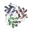
| ||||||||
| Unit cell |
|
-Components
| #1: Protein | Mass: 17190.844 Da / Num. of mol.: 3 Source method: isolated from a genetically manipulated source Source: (gene. exp.) Bos taurus (cattle) / Tissue: RETINA / Organ: EYE / Production host:  Escherichia coli (E. coli) / References: UniProt: P52175, nucleoside-diphosphate kinase Escherichia coli (E. coli) / References: UniProt: P52175, nucleoside-diphosphate kinase#2: Chemical | Cyclic guanosine monophosphate  Mass: 345.205 Da / Num. of mol.: 3 / Source method: obtained synthetically / Formula: C10H12N5O7P Mass: 345.205 Da / Num. of mol.: 3 / Source method: obtained synthetically / Formula: C10H12N5O7P#3: Water | ChemComp-HOH / | Water Mass: 18.015 Da / Num. of mol.: 278 / Source method: isolated from a natural source / Formula: H2O Mass: 18.015 Da / Num. of mol.: 278 / Source method: isolated from a natural source / Formula: H2O |
|---|
-Experimental details
-Experiment
| Experiment | Method: X-RAY DIFFRACTION / Number of used crystals: 1 |
|---|
- Sample preparation
| Crystal | Density Matthews: 3.5 Å3/Da / Density % sol: 65.19 % | ||||||||||||||||||||
|---|---|---|---|---|---|---|---|---|---|---|---|---|---|---|---|---|---|---|---|---|---|
| Crystal grow | pH: 7 / Details: pH 7 | ||||||||||||||||||||
| Crystal grow | *PLUS Method: vapor diffusion, hanging dropDetails: drops were formed by combining equal volumes of protein solution and reservoir solution | ||||||||||||||||||||
| Components of the solutions | *PLUS
|
-Data collection
| Diffraction | Mean temperature: 100 K |
|---|---|
| Diffraction source | Source: ROTATING ANODE / Type: SIEMENS / Wavelength: 1.5418 |
| Detector | Type: SIEMENS HI-STAR / Detector: AREA DETECTOR / Date: Mar 1, 1997 / Details: COLLIMATOR |
| Radiation | Monochromator: GRAPHITE(002) / Monochromatic (M) / Laue (L): M / Scattering type: x-ray |
| Radiation wavelength | Wavelength: 1.5418 Å / Relative weight: 1 |
| Reflection | Resolution: 2.4→20 Å / Num. obs: 30274 / % possible obs: 89 % / Redundancy: 4.6 % / Rsym value: 0.109 / Net I/σ(I): 7.5 |
| Reflection shell | Resolution: 2.4→2.51 Å / Mean I/σ(I) obs: 1.3 / % possible all: 60 |
| Reflection | *PLUS Rmerge(I) obs: 0.109 |
| Reflection shell | *PLUS % possible obs: 60 % |
- Processing
| Software |
| ||||||||||||||||||||||||||||||||||||||||||||||||||
|---|---|---|---|---|---|---|---|---|---|---|---|---|---|---|---|---|---|---|---|---|---|---|---|---|---|---|---|---|---|---|---|---|---|---|---|---|---|---|---|---|---|---|---|---|---|---|---|---|---|---|---|
| Refinement | Method to determine structure: MOLECULAR REPLACEMENT Starting model: PDB ENTRY 1NSK Resolution: 2.4→20 Å / Isotropic thermal model: TNT BCORREL Details: X-PLOR SIMULATED ANNEALING, SLOW COOLING, POSITIONAL AND B-FACTOR REFINEMENT WAS USED WITH 10% OF THE DATA RESERVED FOR R-FREE. R-FREE WENT FROM 0.398 - 0.366 WHILE THE WORKING R WENT TO 0. ...Details: X-PLOR SIMULATED ANNEALING, SLOW COOLING, POSITIONAL AND B-FACTOR REFINEMENT WAS USED WITH 10% OF THE DATA RESERVED FOR R-FREE. R-FREE WENT FROM 0.398 - 0.366 WHILE THE WORKING R WENT TO 0.259. THEN TNT WAS USED FOR THE FINAL REFINEMENT WITHOUT R-FREE.
| ||||||||||||||||||||||||||||||||||||||||||||||||||
| Solvent computation | Bsol: 283.9 Å2 / ksol: 0.809 e/Å3 | ||||||||||||||||||||||||||||||||||||||||||||||||||
| Refinement step | Cycle: LAST / Resolution: 2.4→20 Å
| ||||||||||||||||||||||||||||||||||||||||||||||||||
| Refine LS restraints |
| ||||||||||||||||||||||||||||||||||||||||||||||||||
| Software | *PLUS Name: TNT / Version: 5E / Classification: refinement | ||||||||||||||||||||||||||||||||||||||||||||||||||
| Refinement | *PLUS | ||||||||||||||||||||||||||||||||||||||||||||||||||
| Solvent computation | *PLUS | ||||||||||||||||||||||||||||||||||||||||||||||||||
| Displacement parameters | *PLUS | ||||||||||||||||||||||||||||||||||||||||||||||||||
| Refine LS restraints | *PLUS Type: t_plane_restr / Dev ideal: 0.026 / Weight: 5.8 |
