 Movie
Movie Controller


+ Open data

- Basic information
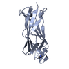
| Entry | Database: PDB / ID: 1bd8 | ||||||
|---|---|---|---|---|---|---|---|
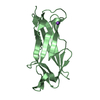
| Title | STRUCTURE OF CDK INHIBITOR P19INK4D | ||||||
 Components Components | P19INK4D CDK4/6 INHIBITOR | ||||||
 Keywords Keywords | TUMOR SUPPRESSOR / CDK4/6 INHIBITOR / ANKYRIN MOTIF | ||||||
| Function / homology |  Function and homology information Function and homology informationcyclin D2-CDK4 complex / autophagic cell death / negative regulation of intrinsic apoptotic signaling pathway in response to DNA damage / cyclin-dependent protein serine/threonine kinase inhibitor activity / response to vitamin D / negative regulation of G1/S transition of mitotic cell cycle / DNA synthesis involved in DNA repair / regulation of G1/S transition of mitotic cell cycle / response to retinoic acid / response to UV ...cyclin D2-CDK4 complex / autophagic cell death / negative regulation of intrinsic apoptotic signaling pathway in response to DNA damage / cyclin-dependent protein serine/threonine kinase inhibitor activity / response to vitamin D / negative regulation of G1/S transition of mitotic cell cycle / DNA synthesis involved in DNA repair / regulation of G1/S transition of mitotic cell cycle / response to retinoic acid / response to UV / sensory perception of sound / negative regulation of cell growth / Oncogene Induced Senescence / Cyclin D associated events in G1 / Senescence-Associated Secretory Phenotype (SASP) / Oxidative Stress Induced Senescence / negative regulation of cell population proliferation / protein kinase binding / nucleoplasm / nucleus / cytoplasm / cytosol Similarity search - Function | ||||||
| Biological species |  Homo sapiens (human) Homo sapiens (human) | ||||||
| Method |  X-RAY DIFFRACTION / MIR / Resolution: 1.8 Å X-RAY DIFFRACTION / MIR / Resolution: 1.8 Å | ||||||
 Authors Authors | Baumgartner, R. / Fernandez-Catalan, C. / Winoto, A. / Huber, R. / Engh, R. / Holak, T.A. | ||||||
 Citation Citation | Journal: Structure / Year: 1998 Title: Structure of human cyclin-dependent kinase inhibitor p19INK4d: comparison to known ankyrin-repeat-containing structures and implications for the dysfunction of tumor suppressor p16INK4a. Authors: Baumgartner, R. / Fernandez-Catalan, C. / Winoto, A. / Huber, R. / Engh, R.A. / Holak, T.A. #1: Journal: Nat.Struct.Biol. / Year: 1998Title: Crystal Structure of the Cdk4/6 Inhibitory Protein P18Ink4C Provides Insights Into Ankyrin-Like Repeat Structure/Function and Tumor-Derived P16Ink4 Mutations Authors: Venkataramani, R. / Swaminathan, K. / Marmorstein, R. | ||||||
| History |
|
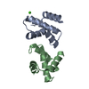
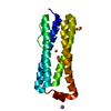
- Structure visualization
| Structure viewer | Molecule: MolmilJmol/JSmol |
|---|
- Downloads & links
-Download
| PDBx/mmCIF format | 1bd8.cif.gz | 45.1 KB | Display | PDBx/mmCIF format |
|---|---|---|---|---|
| PDB format | pdb1bd8.ent.gz | 32.4 KB | Display | PDB format |
| PDBx/mmJSON format | 1bd8.json.gz | Tree view | PDBx/mmJSON format | |
| Others |  Other downloads Other downloads |
-Validation report
| Arichive directory | https://data.pdbj.org/pub/pdb/validation_reports/bd/1bd8ftp://data.pdbj.org/pub/pdb/validation_reports/bd/1bd8 | HTTPS FTP |
|---|
-Related structure data
| Similar structure data |
|---|


-Links
 PDBj
PDBj


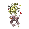
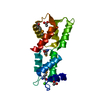
- Assembly
| Deposited unit | 
| ||||||||
|---|---|---|---|---|---|---|---|---|---|
| 1 |
| ||||||||
| Unit cell |
|
-Components
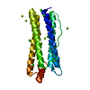
| #1: Protein | Mass: 16627.812 Da / Num. of mol.: 1 Source method: isolated from a genetically manipulated source Source: (gene. exp.) Homo sapiens (human) / Cell line: BL21 / Cellular location: CYTOPLASM / Plasmid: PET-15B / Species (production host): Escherichia coli / Cellular location (production host): CYTOPLASM / Production host:  |
|---|---|
| #2: Water | ChemComp-HOH /  Mass: 18.015 Da / Num. of mol.: 195 / Source method: isolated from a natural source / Formula: H2O Mass: 18.015 Da / Num. of mol.: 195 / Source method: isolated from a natural source / Formula: H2O |
-Experimental details
-Experiment
| Experiment | Method: X-RAY DIFFRACTION / Number of used crystals: 1 |
|---|
- Sample preparation
| Crystal | Density Matthews: 1.86 Å3/Da / Density % sol: 34 % / Description: DATA COLLECTED IN THE OSCILLATION MODE. | ||||||||||||||||||||||||||||||
|---|---|---|---|---|---|---|---|---|---|---|---|---|---|---|---|---|---|---|---|---|---|---|---|---|---|---|---|---|---|---|---|
| Crystal grow | Temperature: 277 K / Method: vapor diffusion, sitting drop / pH: 8.5 Details: PROTEIN WAS CRYSTALLIZED FROM 35% PEG 4000, 200 MM MGSO4 AND 200 MM TRIS/HCL PH 8.5, SITTING DROP METHOD, AT 4 DEG. C., vapor diffusion - sitting drop, temperature 277K | ||||||||||||||||||||||||||||||
| Crystal | *PLUS Density % sol: 35 % | ||||||||||||||||||||||||||||||
| Crystal grow | *PLUS Temperature: 4 ℃ / Method: vapor diffusion, sitting drop | ||||||||||||||||||||||||||||||
| Components of the solutions | *PLUS
|
-Data collection
| Diffraction | Mean temperature: 280 K |
|---|---|
| Diffraction source | Source: ROTATING ANODE / Type: RIGAKU RUH2R / Wavelength: 1.5418 |
| Detector | Type: MARRESEARCH / Detector: IMAGE PLATE / Date: Oct 1, 1997 / Details: MIRRORS |
| Radiation | Monochromator: GRAPHITE(002) / Monochromatic (M) / Laue (L): M / Scattering type: x-ray |
| Radiation wavelength | Wavelength: 1.5418 Å / Relative weight: 1 |
| Reflection | Resolution: 1.8→25 Å / Num. obs: 13017 / % possible obs: 99.9 % / Observed criterion σ(I): 2 / Rmerge(I) obs: 0.073 / Net I/σ(I): 11.7 |
| Reflection | *PLUS % possible obs: 99.4 % |
- Processing
| Software |
| ||||||||||||||||||
|---|---|---|---|---|---|---|---|---|---|---|---|---|---|---|---|---|---|---|---|
| Refinement | Method to determine structure: MIR / Highest resolution: 1.8 Å /
| ||||||||||||||||||
| Refinement step | Cycle: LAST / Highest resolution: 1.8 Å
| ||||||||||||||||||
| Software | *PLUS Name: REFMAC / Classification: refinement | ||||||||||||||||||
| Refinement | *PLUS Rfactor obs: 0.19 | ||||||||||||||||||
| Solvent computation | *PLUS | ||||||||||||||||||
| Displacement parameters | *PLUS | ||||||||||||||||||
| Refine LS restraints | *PLUS
|
