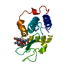
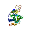
 Movie
Movie Controller


+ Open data

- Basic information
| Entry | Database: PDB / ID: 1bb6 | ||||||
|---|---|---|---|---|---|---|---|
| Title | LYSOZYME COMPLEX WITH 4-METHYL-UMBELLIFERYL CHITOTRIOSE | ||||||
 Components Components | LYSOZYME | ||||||
 Keywords Keywords | HYDROLASE / N-ACETYL-MURAMIDASE / UMBELLIFERONE GLYCOSIDES | ||||||
| Function / homology |  Function and homology informationmetabolic process / lysozyme / lysozyme activity / killing of cells of another organism / defense response to bacterium / extracellular space Function and homology informationmetabolic process / lysozyme / lysozyme activity / killing of cells of another organism / defense response to bacterium / extracellular spaceSimilarity search - Function | ||||||
| Biological species |  Oncorhynchus mykiss (rainbow trout) Oncorhynchus mykiss (rainbow trout) | ||||||
| Method | X-RAY DIFFRACTION / PHASES FOR NATIVE STRUCTURE / Resolution: 2 Å | ||||||
 Authors Authors | Vollan, V.B. / Hough, E. / Karlsen, S. | ||||||
 Citation Citation | Journal: Acta Crystallogr.,Sect.D / Year: 1999 Title: Structural studies on the binding of 4-methylumbelliferone glycosides of chitin to rainbow trout lysozyme. Authors: Vollan, V.B. / Hough, E. / Karlsen, S. #1: Journal: Acta Crystallogr.,Sect.D / Year: 1995Title: Crystal Structures of Three Complexes between Chito-Oligosaccharides and Lysozyme from the Rainbow Trout. How Distorted is the Nag Sugar in Site D? Authors: Karlsen, S. / Hough, E. #2: Journal: Acta Crystallogr.,Sect.D / Year: 1995Title: Refined Crystal Structure of Lysozyme from the Rainbow Trout (Oncorhynchus Mykiss) Authors: Karlsen, S. / Eliassen, B.E. / Hansen, L.K. / Larsen, R.L. / Riise, B.W. / Smalas, A.O. / Hough, E. / Grinde, B. | ||||||
| History |
|
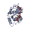
- Structure visualization
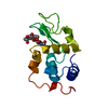
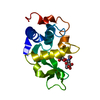
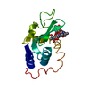
| Structure viewer | Molecule: MolmilJmol/JSmol |
|---|
- Downloads & links
-Download
| PDBx/mmCIF format | 1bb6.cif.gz | 37.9 KB | Display | PDBx/mmCIF format |
|---|---|---|---|---|
| PDB format | pdb1bb6.ent.gz | 29.6 KB | Display | PDB format |
| PDBx/mmJSON format | 1bb6.json.gz | Tree view | PDBx/mmJSON format | |
| Others |  Other downloads Other downloads |
-Validation report
| Arichive directory | https://data.pdbj.org/pub/pdb/validation_reports/bb/1bb6ftp://data.pdbj.org/pub/pdb/validation_reports/bb/1bb6 | HTTPS FTP |
|---|
-Related structure data


-Links
 PDBj
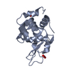
PDBj- Assembly
| Deposited unit | 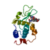
| ||||||||
|---|---|---|---|---|---|---|---|---|---|
| 1 |
| ||||||||
| Unit cell |
|
-Components
| #1: Protein | / N-ACETYL-MURAMIDASE Mass: 14303.068 Da / Num. of mol.: 1 / Source method: isolated from a natural source / Source: (natural) Oncorhynchus mykiss (rainbow trout) / Organ: KIDNEY / References: UniProt: P11941, lysozyme |
|---|---|
| #2: Chemical | ChemComp-UMG / 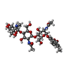  Mass: 785.746 Da / Num. of mol.: 1 / Source method: obtained synthetically / Formula: C34H47N3O18 Mass: 785.746 Da / Num. of mol.: 1 / Source method: obtained synthetically / Formula: C34H47N3O18 |
| #3: Water | ChemComp-HOH / Water Mass: 18.015 Da / Num. of mol.: 142 / Source method: isolated from a natural source / Formula: H2O Mass: 18.015 Da / Num. of mol.: 142 / Source method: isolated from a natural source / Formula: H2O |
-Experimental details
-Experiment
| Experiment | Method: X-RAY DIFFRACTION / Number of used crystals: 1 |
|---|
- Sample preparation
| Crystal | Density Matthews: 3.25 Å3/Da / Density % sol: 62 % | ||||||||||||||||||||||||||||||||||||
|---|---|---|---|---|---|---|---|---|---|---|---|---|---|---|---|---|---|---|---|---|---|---|---|---|---|---|---|---|---|---|---|---|---|---|---|---|---|
| Crystal grow | pH: 10 / Details: pH 10.0 | ||||||||||||||||||||||||||||||||||||
| Crystal | *PLUS | ||||||||||||||||||||||||||||||||||||
| Crystal grow | *PLUS Temperature: 277 K / Method: vapor diffusion, hanging dropDetails: Karlsen, S., (1995) Acta Crystallogr.,Sect.D, 51, 354. PH range low: 10 / PH range high: 4 | ||||||||||||||||||||||||||||||||||||
| Components of the solutions | *PLUS
|
-Data collection
| Diffraction | Mean temperature: 298 K |
|---|---|
| Diffraction source | Source: ROTATING ANODE / Type: ENRAF-NONIUS FR571 / Wavelength: 1.5418 |
| Detector | Type: ENRAF-NONIUS FAST / Detector: DIFFRACTOMETER / Date: Mar 1, 1996 |
| Radiation | Monochromator: GRAPHITE(002) / Monochromatic (M) / Laue (L): M / Scattering type: x-ray |
| Radiation wavelength | Wavelength: 1.5418 Å / Relative weight: 1 |
| Reflection | Resolution: 2→28 Å / Num. obs: 11445 / % possible obs: 89.5 % / Observed criterion σ(I): 1 / Redundancy: 1.8 % / Rmerge(I) obs: 0.01 / Net I/σ(I): 5.9 |
| Reflection shell | Resolution: 2→2.1 Å / Redundancy: 1.2 % / Mean I/σ(I) obs: 1.8 / % possible all: 42.7 |
| Reflection | *PLUS Num. measured all: 20088 / Rmerge(I) obs: 0.105 |
| Reflection shell | *PLUS % possible obs: 42.7 % |
- Processing
| Software |
| ||||||||||||||||||||||||||||||||||||||||||||||||||||||||||||||||||||||||||||||||||||
|---|---|---|---|---|---|---|---|---|---|---|---|---|---|---|---|---|---|---|---|---|---|---|---|---|---|---|---|---|---|---|---|---|---|---|---|---|---|---|---|---|---|---|---|---|---|---|---|---|---|---|---|---|---|---|---|---|---|---|---|---|---|---|---|---|---|---|---|---|---|---|---|---|---|---|---|---|---|---|---|---|---|---|---|---|---|
| Refinement | Method to determine structure: PHASES FOR NATIVE STRUCTURE / Resolution: 2→8 Å / Cross valid method: THROUGHOUT / σ(F): 1
| ||||||||||||||||||||||||||||||||||||||||||||||||||||||||||||||||||||||||||||||||||||
| Displacement parameters | Biso mean: 18.87 Å2 | ||||||||||||||||||||||||||||||||||||||||||||||||||||||||||||||||||||||||||||||||||||
| Refine analyze | Luzzati coordinate error obs: 0.25 Å | ||||||||||||||||||||||||||||||||||||||||||||||||||||||||||||||||||||||||||||||||||||
| Refinement step | Cycle: LAST / Resolution: 2→8 Å
| ||||||||||||||||||||||||||||||||||||||||||||||||||||||||||||||||||||||||||||||||||||
| Refine LS restraints |
| ||||||||||||||||||||||||||||||||||||||||||||||||||||||||||||||||||||||||||||||||||||
| Software | *PLUS Name: PROLSQ / Classification: refinement | ||||||||||||||||||||||||||||||||||||||||||||||||||||||||||||||||||||||||||||||||||||
| Refinement | *PLUS Rfactor obs: 0.211 | ||||||||||||||||||||||||||||||||||||||||||||||||||||||||||||||||||||||||||||||||||||
| Solvent computation | *PLUS | ||||||||||||||||||||||||||||||||||||||||||||||||||||||||||||||||||||||||||||||||||||
| Displacement parameters | *PLUS |
