 Movie
Movie Controller


+ Open data

- Basic information
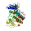
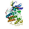
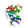
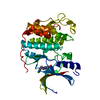
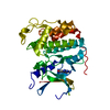
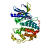
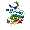
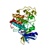
| Entry | Database: PDB / ID: 1b38 | ||||||
|---|---|---|---|---|---|---|---|
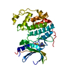
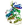
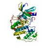
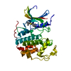
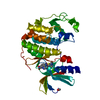
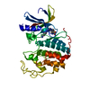
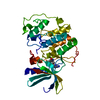
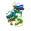
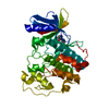
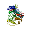
| Title | HUMAN CYCLIN-DEPENDENT KINASE 2 | ||||||
 Components Components | PROTEIN (CELL DIVISION PROTEIN KINASE 2) | ||||||
 Keywords Keywords | TRANSFERASE / PROTEIN KINASE / SERINE/THREONINE PROTEIN KINASE / ATP-BINDING / CELL CYCLE / CELL DIVISION / MITOSIS / PHOSPHORYLATION | ||||||
| Function / homology |  Function and homology information Function and homology informationReplication initiator protein RctB, central region / RctB, helix turn helix domain / Vibrionales, replication initiator protein RctB, central region / RctB helix turn helix domain / Transferase(Phosphotransferase) domain 1 / Transferase(Phosphotransferase); domain 1 / Phosphorylase Kinase; domain 1 / Phosphorylase Kinase; domain 1 / Protein kinase domain / 2-Layer Sandwich ...Replication initiator protein RctB, central region / RctB, helix turn helix domain / Vibrionales, replication initiator protein RctB, central region / RctB helix turn helix domain / Transferase(Phosphotransferase) domain 1 / Transferase(Phosphotransferase); domain 1 / Phosphorylase Kinase; domain 1 / Phosphorylase Kinase; domain 1 / Protein kinase domain / 2-Layer Sandwich / Orthogonal Bundle / Mainly Alpha / Alpha Beta Similarity search - Domain/homology | ||||||
| Biological species |  Homo sapiens (human) Homo sapiens (human) | ||||||
| Method |  X-RAY DIFFRACTION / SYNCHROTRON / OTHER / Resolution: 2 Å X-RAY DIFFRACTION / SYNCHROTRON / OTHER / Resolution: 2 Å | ||||||
 Authors Authors | Brown, N.R. / Noble, M.E.M. / Lawrie, A.M. / Morris, M.C. / Tunnah, P. / Divita, G. / Johnson, L.N. / Endicott, J.A. | ||||||
 Citation Citation | Journal: J.Biol.Chem. / Year: 1999 Title: Effects of phosphorylation of threonine 160 on cyclin-dependent kinase 2 structure and activity. Authors: Brown, N.R. / Noble, M.E. / Lawrie, A.M. / Morris, M.C. / Tunnah, P. / Divita, G. / Johnson, L.N. / Endicott, J.A. #1: Journal: Proteins / Year: 1995Title: Multiple Modes of Ligand Recognition: Crystal Structures of Cyclin-Dependent Protein Kinase 2 in Complex with ATP and Two Inhibitors, Olomoucine and Isopentenyladenine Authors: Schulze-Gahmen, U. / Brandsen, J. / Jones, H.D. / Morgan, D.O. / Meijer, L. / Vesely, J. / Kim, S.H. | ||||||
| History |
|
- Structure visualization
| Structure viewer | Molecule: MolmilJmol/JSmol |
|---|
- Downloads & links
-Download
| PDBx/mmCIF format | 1b38.cif.gz | 78.6 KB | Display | PDBx/mmCIF format |
|---|---|---|---|---|
| PDB format | pdb1b38.ent.gz | 57.2 KB | Display | PDB format |
| PDBx/mmJSON format | 1b38.json.gz | Tree view | PDBx/mmJSON format | |
| Others |  Other downloads Other downloads |
-Validation report
| Summary document | 1b38_validation.pdf.gz | 763.5 KB | Display | wwPDB validaton report |
|---|---|---|---|---|
| Full document | 1b38_full_validation.pdf.gz | 775.1 KB | Display | |
| Data in XML | 1b38_validation.xml.gz | 16.8 KB | Display | |
| Data in CIF | 1b38_validation.cif.gz | 24.1 KB | Display | |
| Arichive directory | https://data.pdbj.org/pub/pdb/validation_reports/b3/1b38ftp://data.pdbj.org/pub/pdb/validation_reports/b3/1b38 | HTTPS FTP |
-Related structure data


-Links
 PDBj
PDBj
- Assembly
| Deposited unit | 
| ||||||||
|---|---|---|---|---|---|---|---|---|---|
| 1 |
| ||||||||
| Unit cell |
|
-Components
| #1: Protein | Mass: 34002.527 Da / Num. of mol.: 1 / Fragment: INTACT Source method: isolated from a genetically manipulated source Source: (gene. exp.) Homo sapiens (human) / Plasmid: BACULOVIRUS / Cell line (production host): SF9 / Production host:   Spodoptera frugiperda (fall armyworm) / References: UniProt: P24941, EC: 2.7.1.37 Spodoptera frugiperda (fall armyworm) / References: UniProt: P24941, EC: 2.7.1.37 |
|---|---|
| #2: Chemical | ChemComp-MG /   Mass: 24.305 Da / Num. of mol.: 1 / Source method: obtained synthetically / Formula: Mg Mass: 24.305 Da / Num. of mol.: 1 / Source method: obtained synthetically / Formula: Mg |
| #3: Chemical | ChemComp-ATP /   Mass: 507.181 Da / Num. of mol.: 1 / Source method: obtained synthetically / Formula: C10H16N5O13P3 / Comment: ATP, energy-carrying molecule*YM Mass: 507.181 Da / Num. of mol.: 1 / Source method: obtained synthetically / Formula: C10H16N5O13P3 / Comment: ATP, energy-carrying molecule*YM |
| #4: Water | ChemComp-HOH /  Mass: 18.015 Da / Num. of mol.: 206 / Source method: isolated from a natural source / Formula: H2O Mass: 18.015 Da / Num. of mol.: 206 / Source method: isolated from a natural source / Formula: H2O |
-Experimental details
-Experiment
| Experiment | Method: X-RAY DIFFRACTION / Number of used crystals: 1 |
|---|
- Sample preparation
| Crystal | Density Matthews: 2.01 Å3/Da / Density % sol: 45 % | ||||||||||||||||||||||||||||||||||||||||||
|---|---|---|---|---|---|---|---|---|---|---|---|---|---|---|---|---|---|---|---|---|---|---|---|---|---|---|---|---|---|---|---|---|---|---|---|---|---|---|---|---|---|---|---|
| Crystal grow | pH: 7.4 Details: PROTEIN AT 10 MG/ML IN 10MM HEPES/HCL PH7.4, 15MM NACL WELL BUFFER CONTAINING 50MM AMMONIUM ACETATE, 12% PEG 3350, 100MM HEPES/HCL PH 7.4 | ||||||||||||||||||||||||||||||||||||||||||
| Crystal grow | *PLUS Method: vapor diffusion, hanging drop | ||||||||||||||||||||||||||||||||||||||||||
| Components of the solutions | *PLUS
|
-Data collection
| Diffraction | Mean temperature: 100 K |
|---|---|
| Diffraction source | Source: SYNCHROTRON / Site: SRS  / Beamline: PX9.5 / Wavelength: 1.5418 / Beamline: PX9.5 / Wavelength: 1.5418 |
| Detector | Type: MAR scanner 300 mm plate / Detector: IMAGE PLATE / Date: May 21, 1998 / Details: MIRROR |
| Radiation | Monochromator: DOUBLE CRYSTAL / Protocol: SINGLE WAVELENGTH / Monochromatic (M) / Laue (L): M / Scattering type: x-ray |
| Radiation wavelength | Wavelength: 1.5418 Å / Relative weight: 1 |
| Reflection | Resolution: 2→20 Å / Num. obs: 18076 / % possible obs: 95.2 % / Observed criterion σ(I): 0 / Redundancy: 3.9 % / Rmerge(I) obs: 0.092 / Net I/σ(I): 11 |
| Reflection shell | Resolution: 2→2.1 Å / Rmerge(I) obs: 0.39 / Mean I/σ(I) obs: 2.26 / % possible all: 96.8 |
| Reflection | *PLUS Num. measured all: 70729 |
| Reflection shell | *PLUS % possible obs: 96.8 % |
- Processing
| Software |
| ||||||||||||||||||||||||||||||||||||||||||||||||||||||||||||||||||||||||||||||||||||
|---|---|---|---|---|---|---|---|---|---|---|---|---|---|---|---|---|---|---|---|---|---|---|---|---|---|---|---|---|---|---|---|---|---|---|---|---|---|---|---|---|---|---|---|---|---|---|---|---|---|---|---|---|---|---|---|---|---|---|---|---|---|---|---|---|---|---|---|---|---|---|---|---|---|---|---|---|---|---|---|---|---|---|---|---|---|
| Refinement | Method to determine structure: OTHER Starting model: UNPUBLISHED Resolution: 2→20 Å / Cross valid method: FREE-R / σ(F): 0
| ||||||||||||||||||||||||||||||||||||||||||||||||||||||||||||||||||||||||||||||||||||
| Refinement step | Cycle: LAST / Resolution: 2→20 Å
| ||||||||||||||||||||||||||||||||||||||||||||||||||||||||||||||||||||||||||||||||||||
| Refine LS restraints |
|
