 Movie
Movie Controller

[English] 日本語
 Yorodumi
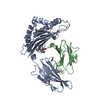
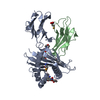
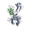
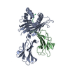
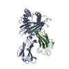
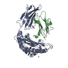
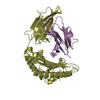
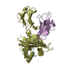
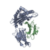
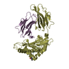
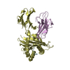
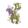
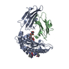
- PDB-1b0g: CLASS I HISTOCOMPATIBILITY ANTIGEN (HLA-A2.1)/BETA 2-MICROGLOBULI... -
+ Open data

- Basic information
| Entry | Database: PDB / ID: 1b0g | |||||||||
|---|---|---|---|---|---|---|---|---|---|---|
| Title | CLASS I HISTOCOMPATIBILITY ANTIGEN (HLA-A2.1)/BETA 2-MICROGLOBULIN/PEPTIDE P1049 COMPLEX | |||||||||
 Components Components |
| |||||||||
 Keywords Keywords | HISTOCOMPATIBILITY ANTIGEN / CLASS I HISTOCOMPATIBILITY ANTIGEN (HLA-A2.1)-BETA 2-MICROGLOBULIN-PEPTIDE P1049 COMPLEX | |||||||||
| Function / homology |  Function and homology information Function and homology informationEMC complex / protein insertion into ER membrane by stop-transfer membrane-anchor sequence / tail-anchored membrane protein insertion into ER membrane / positive regulation of memory T cell activation / T cell mediated cytotoxicity directed against tumor cell target / TAP complex binding / positive regulation of CD8-positive, alpha-beta T cell activation / Golgi medial cisterna / CD8-positive, alpha-beta T cell activation / positive regulation of CD8-positive, alpha-beta T cell proliferation ...EMC complex / protein insertion into ER membrane by stop-transfer membrane-anchor sequence / tail-anchored membrane protein insertion into ER membrane / positive regulation of memory T cell activation / T cell mediated cytotoxicity directed against tumor cell target / TAP complex binding / positive regulation of CD8-positive, alpha-beta T cell activation / Golgi medial cisterna / CD8-positive, alpha-beta T cell activation / positive regulation of CD8-positive, alpha-beta T cell proliferation / CD8 receptor binding / antigen processing and presentation of exogenous peptide antigen via MHC class I / antigen processing and presentation of endogenous peptide antigen via MHC class I via ER pathway, TAP-dependent / beta-2-microglobulin binding / endoplasmic reticulum exit site / TAP binding / protection from natural killer cell mediated cytotoxicity / antigen processing and presentation of endogenous peptide antigen via MHC class Ib / antigen processing and presentation of endogenous peptide antigen via MHC class I via ER pathway, TAP-independent / detection of bacterium / T cell receptor binding / early endosome lumen / Nef mediated downregulation of MHC class I complex cell surface expression / DAP12 interactions / Endosomal/Vacuolar pathway / Antigen Presentation: Folding, assembly and peptide loading of class I MHC / negative regulation of iron ion transport / lumenal side of endoplasmic reticulum membrane / T cell mediated cytotoxicity / cellular response to iron(III) ion / negative regulation of forebrain neuron differentiation / antigen processing and presentation of exogenous protein antigen via MHC class Ib, TAP-dependent / ER to Golgi transport vesicle membrane / peptide antigen assembly with MHC class I protein complex / transferrin transport / regulation of iron ion transport / regulation of erythrocyte differentiation / negative regulation of receptor-mediated endocytosis / HFE-transferrin receptor complex / response to molecule of bacterial origin / MHC class I peptide loading complex / cellular response to iron ion / positive regulation of T cell cytokine production / antigen processing and presentation of endogenous peptide antigen via MHC class I / MHC class I protein complex / peptide antigen assembly with MHC class II protein complex / negative regulation of neurogenesis / positive regulation of receptor-mediated endocytosis / cellular response to nicotine / MHC class II protein complex / positive regulation of T cell mediated cytotoxicity / multicellular organismal-level iron ion homeostasis / specific granule lumen / peptide antigen binding / antigen processing and presentation of exogenous peptide antigen via MHC class II / positive regulation of type II interferon production / phagocytic vesicle membrane / positive regulation of immune response / recycling endosome membrane / Interferon gamma signaling / positive regulation of T cell activation / Immunoregulatory interactions between a Lymphoid and a non-Lymphoid cell / negative regulation of epithelial cell proliferation / Interferon alpha/beta signaling / Modulation by Mtb of host immune system / sensory perception of smell / positive regulation of cellular senescence / tertiary granule lumen / DAP12 signaling / MHC class II protein complex binding / T cell differentiation in thymus / T cell receptor signaling pathway / late endosome membrane / negative regulation of neuron projection development / antibacterial humoral response / E3 ubiquitin ligases ubiquitinate target proteins / ER-Phagosome pathway / carbohydrate binding / protein refolding / early endosome membrane / amyloid fibril formation / protein homotetramerization / intracellular iron ion homeostasis / learning or memory / defense response to Gram-positive bacterium / immune response / endoplasmic reticulum lumen / Amyloid fiber formation / Golgi membrane / signaling receptor binding / external side of plasma membrane / innate immune response / lysosomal membrane / focal adhesion / Neutrophil degranulation / endoplasmic reticulum membrane / SARS-CoV-2 activates/modulates innate and adaptive immune responses / structural molecule activity / cell surface / endoplasmic reticulum Similarity search - Function | |||||||||
| Biological species |  Homo sapiens (human) Homo sapiens (human) | |||||||||
| Method |  X-RAY DIFFRACTION / MOLECULAR REPLACEMENT / Resolution: 2.5 Å X-RAY DIFFRACTION / MOLECULAR REPLACEMENT / Resolution: 2.5 Å | |||||||||
 Authors Authors | Zhao, R. / Collins, E.J. | |||||||||
 Citation Citation | Journal: J.Exp.Med. / Year: 1999 Title: Structural evidence of T cell xeno-reactivity in the absence of molecular mimicry. Authors: Zhao, R. / Loftus, D.J. / Appella, E. / Collins, E.J. | |||||||||
| History |
|
- Structure visualization
| Structure viewer | Molecule: MolmilJmol/JSmol |
|---|
- Downloads & links
-Download
| PDBx/mmCIF format | 1b0g.cif.gz | 163.8 KB | Display | PDBx/mmCIF format |
|---|---|---|---|---|
| PDB format | pdb1b0g.ent.gz | 131 KB | Display | PDB format |
| PDBx/mmJSON format | 1b0g.json.gz | Tree view | PDBx/mmJSON format | |
| Others |  Other downloads Other downloads |
-Validation report
| Arichive directory | https://data.pdbj.org/pub/pdb/validation_reports/b0/1b0gftp://data.pdbj.org/pub/pdb/validation_reports/b0/1b0g | HTTPS FTP |
|---|
-Related structure data
| Related structure data | 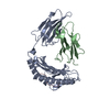 1bz9C 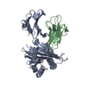 1hhjS S: Starting model for refinement C: citing same article ( |
|---|---|
| Similar structure data |


-Links
 PDBj
PDBj


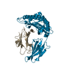
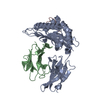
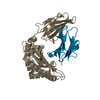
- Assembly
| Deposited unit | 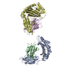
| ||||||||||
|---|---|---|---|---|---|---|---|---|---|---|---|
| 1 | 
| ||||||||||
| 2 | 
| ||||||||||
| Unit cell |
| ||||||||||
| Noncrystallographic symmetry (NCS) | NCS oper: (Code: given Matrix: (-0.914367, 0.404851, -0.005413), Vector: |
-Components
| #1: Protein | Mass: 31854.203 Da / Num. of mol.: 2 Source method: isolated from a genetically manipulated source Source: (gene. exp.) Homo sapiens (human) / Cellular location: CELL SURFACE / Plasmid: PLM1 / Cellular location (production host): CYTOPLASM / Production host:  #2: Protein | Mass: 11879.356 Da / Num. of mol.: 2 Source method: isolated from a genetically manipulated source Source: (gene. exp.) Homo sapiens (human) / Plasmid: PLM1 / Cellular location (production host): CYTOPLASM / Production host: #3: Protein/peptide | Mass: 1049.263 Da / Num. of mol.: 2 Source method: isolated from a genetically manipulated source References: UniProt: Q9NPA0*PLUS #4: Water | ChemComp-HOH / |  Mass: 18.015 Da / Num. of mol.: 50 / Source method: isolated from a natural source / Formula: H2O Mass: 18.015 Da / Num. of mol.: 50 / Source method: isolated from a natural source / Formula: H2OHas protein modification | Y | |
|---|
-Experimental details
-Experiment
| Experiment | Method: X-RAY DIFFRACTION / Number of used crystals: 1 |
|---|
- Sample preparation
| Crystal | Density Matthews: 2.5 Å3/Da / Density % sol: 50.8 % | ||||||||||||||||||||||||||||||||||||||||||
|---|---|---|---|---|---|---|---|---|---|---|---|---|---|---|---|---|---|---|---|---|---|---|---|---|---|---|---|---|---|---|---|---|---|---|---|---|---|---|---|---|---|---|---|
| Crystal grow | Method: vapor diffusion, hanging drop / pH: 6.5 Details: PROTEIN WAS CRYSTALLIZED IN THE HANGING DROP VAPOR DIFFUSION METHOD. THE RESERVOIR SOLUTION CONTAINED 12-16% PEG6000, 6% DIOXANE IN 25MM MES BUFFER, PH6.5. THE HANGING DROP WAS A 1:1 MIXTURE ...Details: PROTEIN WAS CRYSTALLIZED IN THE HANGING DROP VAPOR DIFFUSION METHOD. THE RESERVOIR SOLUTION CONTAINED 12-16% PEG6000, 6% DIOXANE IN 25MM MES BUFFER, PH6.5. THE HANGING DROP WAS A 1:1 MIXTURE OF THE RESERVOIR SOLUTION AND THE PROTEIN SOLUTION WHICH CONTAINED 10MG/ML PROTEIN IN 25MM MES BUFFER, PH6.5., vapor diffusion - hanging drop | ||||||||||||||||||||||||||||||||||||||||||
| Components of the solutions |
| ||||||||||||||||||||||||||||||||||||||||||
| Crystal grow | *PLUS Method: vapor diffusion, hanging drop | ||||||||||||||||||||||||||||||||||||||||||
| Components of the solutions | *PLUS
|
-Data collection
| Diffraction | Mean temperature: 100 K |
|---|---|
| Diffraction source | Source: ROTATING ANODE / Type: RIGAKU RU200 / Wavelength: 1.5418 |
| Detector | Type: RIGAKU RAXIS IIC / Detector: IMAGE PLATE / Date: May 15, 1997 / Details: MIRRORS |
| Radiation | Monochromator: NI FILTER / Protocol: MONOCHROMATIC / Monochromatic (M) / Laue (L): M / Scattering type: x-ray |
| Radiation wavelength | Wavelength: 1.5418 Å / Relative weight: 1 |
| Reflection | Resolution: 2.5→30 Å / Num. all: 29804 / Num. obs: 29804 / % possible obs: 90.9 % / Observed criterion σ(I): 0 / Redundancy: 2.6 % / Biso Wilson estimate: 23.42 Å2 / Rmerge(I) obs: 0.083 / Net I/σ(I): 16.6 |
| Reflection shell | Resolution: 2.5→2.59 Å / Redundancy: 1.8 % / Rmerge(I) obs: 0.176 / Mean I/σ(I) obs: 6.5 / % possible all: 70.8 |
| Reflection | *PLUS Num. measured all: 77499 |
| Reflection shell | *PLUS Highest resolution: 2.5 Å / % possible obs: 70.8 % |
- Processing
| Software |
| ||||||||||||||||||||||||||||||||||||||||||||||||||||||||||||||||||||||||||||||||||||
|---|---|---|---|---|---|---|---|---|---|---|---|---|---|---|---|---|---|---|---|---|---|---|---|---|---|---|---|---|---|---|---|---|---|---|---|---|---|---|---|---|---|---|---|---|---|---|---|---|---|---|---|---|---|---|---|---|---|---|---|---|---|---|---|---|---|---|---|---|---|---|---|---|---|---|---|---|---|---|---|---|---|---|---|---|---|
| Refinement | Method to determine structure: MOLECULAR REPLACEMENT Starting model: PDB ENTRY 1HHJ Resolution: 2.5→30 Å / SU ML: 0.32 / Cross valid method: THROUGHOUT / σ(F): 0 / ESU R: 3.44 / ESU R Free: 0.39
| ||||||||||||||||||||||||||||||||||||||||||||||||||||||||||||||||||||||||||||||||||||
| Displacement parameters | Biso mean: 37.06 Å2
| ||||||||||||||||||||||||||||||||||||||||||||||||||||||||||||||||||||||||||||||||||||
| Refinement step | Cycle: LAST / Resolution: 2.5→30 Å
| ||||||||||||||||||||||||||||||||||||||||||||||||||||||||||||||||||||||||||||||||||||
| Refine LS restraints |
| ||||||||||||||||||||||||||||||||||||||||||||||||||||||||||||||||||||||||||||||||||||
| Refinement | *PLUS Lowest resolution: 30 Å | ||||||||||||||||||||||||||||||||||||||||||||||||||||||||||||||||||||||||||||||||||||
| Solvent computation | *PLUS | ||||||||||||||||||||||||||||||||||||||||||||||||||||||||||||||||||||||||||||||||||||
| Displacement parameters | *PLUS | ||||||||||||||||||||||||||||||||||||||||||||||||||||||||||||||||||||||||||||||||||||
| Refine LS restraints | *PLUS
|
