 Movie
Movie Controller


+ Open data

- Basic information
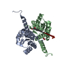
| Entry | Database: PDB / ID: 1aw9 | ||||||
|---|---|---|---|---|---|---|---|
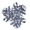
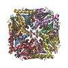
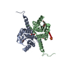
| Title | STRUCTURE OF GLUTATHIONE S-TRANSFERASE III IN APO FORM | ||||||
 Components Components | GLUTATHIONE S-TRANSFERASE III | ||||||
 Keywords Keywords | TRANSFERASE / HERBICIDE DETOXIFICATION | ||||||
| Function / homology |  Function and homology information Function and homology informationresponse to herbicide / glutathione transferase / glutathione transferase activity / glutathione metabolic process / protein homodimerization activity / protein-containing complexSimilarity search - Function | ||||||
| Biological species |  Zea mays (maize) Zea mays (maize) | ||||||
| Method | X-RAY DIFFRACTION / Resolution: 2.2 Å | ||||||
 Authors Authors | Neuefeind, T. / Huber, R. / Reinemer, P. / Knaeblein, J. | ||||||
 Citation Citation | Journal: J.Mol.Biol. / Year: 1997 Title: Cloning, sequencing, crystallization and X-ray structure of glutathione S-transferase-III from Zea mays var. mutin: a leading enzyme in detoxification of maize herbicides. Authors: Neuefeind, T. / Huber, R. / Reinemer, P. / Knablein, J. / Prade, L. / Mann, K. / Bieseler, B. #1: Journal: J.Mol.Biol. / Year: 1997Title: Crystal Structure of Herbicide-Detoxifying Maize Glutathione S-Transferase-I in Complex with Lactoylglutathione: Evidence for an Induced-Fit Mechanism Authors: Neuefeind, T. / Huber, R. / Dasenbrock, H. / Prade, L. / Bieseler, B. | ||||||
| History |
|
- Structure visualization
| Structure viewer | Molecule: MolmilJmol/JSmol |
|---|
- Downloads & links
-Download
| PDBx/mmCIF format | 1aw9.cif.gz | 70.4 KB | Display | PDBx/mmCIF format |
|---|---|---|---|---|
| PDB format | pdb1aw9.ent.gz | 53.2 KB | Display | PDB format |
| PDBx/mmJSON format | 1aw9.json.gz | Tree view | PDBx/mmJSON format | |
| Others |  Other downloads Other downloads |
-Validation report
| Arichive directory | https://data.pdbj.org/pub/pdb/validation_reports/aw/1aw9ftp://data.pdbj.org/pub/pdb/validation_reports/aw/1aw9 | HTTPS FTP |
|---|
-Related structure data
| Similar structure data |
|---|


-Links
 PDBj
PDBj

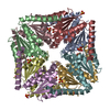
- Assembly
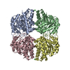
| Deposited unit | 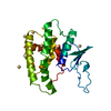
| ||||||||
|---|---|---|---|---|---|---|---|---|---|
| 1 | x 6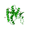
| ||||||||
| Unit cell |
|
-Components
| #1: Protein | Mass: 23456.945 Da / Num. of mol.: 1 Source method: isolated from a genetically manipulated source Details: APO FORM / Source: (gene. exp.) Zea mays (maize) / Cell line: 293 / Variant: MUTIN / Plasmid: PET3A / Production host:  Escherichia coli (E. coli) / Strain (production host): 293 / References: UniProt: Q9ZP62, glutathione transferase Escherichia coli (E. coli) / Strain (production host): 293 / References: UniProt: Q9ZP62, glutathione transferase | ||
|---|---|---|---|
| #2: Chemical | ChemComp-CD /   Mass: 112.411 Da / Num. of mol.: 5 / Source method: obtained synthetically / Formula: Cd Mass: 112.411 Da / Num. of mol.: 5 / Source method: obtained synthetically / Formula: Cd#3: Water | ChemComp-HOH / | Water Mass: 18.015 Da / Num. of mol.: 161 / Source method: isolated from a natural source / Formula: H2O Mass: 18.015 Da / Num. of mol.: 161 / Source method: isolated from a natural source / Formula: H2O |
-Experimental details
-Experiment
| Experiment | Method: X-RAY DIFFRACTION / Number of used crystals: 1 |
|---|
- Sample preparation
| Crystal | Density Matthews: 3.42 Å3/Da / Density % sol: 64.05 % | ||||||||||||||||||||||||||||||||||||||||||||||||||||||||
|---|---|---|---|---|---|---|---|---|---|---|---|---|---|---|---|---|---|---|---|---|---|---|---|---|---|---|---|---|---|---|---|---|---|---|---|---|---|---|---|---|---|---|---|---|---|---|---|---|---|---|---|---|---|---|---|---|---|
| Crystal grow | pH: 7.5 / Details: pH 7.5 | ||||||||||||||||||||||||||||||||||||||||||||||||||||||||
| Crystal grow | *PLUS Method: vapor diffusion, hanging drop / pH: 7 | ||||||||||||||||||||||||||||||||||||||||||||||||||||||||
| Components of the solutions | *PLUS
|
-Data collection
| Diffraction | Mean temperature: 290 K |
|---|---|
| Diffraction source | Source: ROTATING ANODE / Type: RIGAKU RUH2R / Wavelength: 1.5418 |
| Detector | Type: MARRESEARCH / Detector: IMAGE PLATE / Date: Mar 1, 1995 |
| Radiation | Monochromatic (M) / Laue (L): M / Scattering type: x-ray |
| Radiation wavelength | Wavelength: 1.5418 Å / Relative weight: 1 |
| Reflection | Num. obs: 15788 / % possible obs: 99.5 % / Redundancy: 9.5 % / Rmerge(I) obs: 0.087 |
| Reflection shell | Highest resolution: 1.97 Å |
| Reflection | *PLUS Highest resolution: 2.2 Å |
- Processing
| Software |
| ||||||||||||||||||||||||||||||||||||||||||||||||||||||||||||
|---|---|---|---|---|---|---|---|---|---|---|---|---|---|---|---|---|---|---|---|---|---|---|---|---|---|---|---|---|---|---|---|---|---|---|---|---|---|---|---|---|---|---|---|---|---|---|---|---|---|---|---|---|---|---|---|---|---|---|---|---|---|
| Refinement | Resolution: 2.2→8 Å /
| ||||||||||||||||||||||||||||||||||||||||||||||||||||||||||||
| Displacement parameters | Biso mean: 161 Å2 | ||||||||||||||||||||||||||||||||||||||||||||||||||||||||||||
| Refinement step | Cycle: LAST / Resolution: 2.2→8 Å
| ||||||||||||||||||||||||||||||||||||||||||||||||||||||||||||
| Refine LS restraints |
| ||||||||||||||||||||||||||||||||||||||||||||||||||||||||||||
| Software | *PLUS Name: X-PLOR / Classification: refinement | ||||||||||||||||||||||||||||||||||||||||||||||||||||||||||||
| Refinement | *PLUS Rfactor Rfree: 0.224 | ||||||||||||||||||||||||||||||||||||||||||||||||||||||||||||
| Solvent computation | *PLUS | ||||||||||||||||||||||||||||||||||||||||||||||||||||||||||||
| Displacement parameters | *PLUS | ||||||||||||||||||||||||||||||||||||||||||||||||||||||||||||
| Refine LS restraints | *PLUS
| ||||||||||||||||||||||||||||||||||||||||||||||||||||||||||||
| LS refinement shell | *PLUS Highest resolution: 2 Å / Lowest resolution: 2.22 Å / Rfactor obs: 0.298 |
