 Movie
Movie Controller


+ Open data

- Basic information
| Entry | Database: PDB / ID: 1au1 | ||||||||||||
|---|---|---|---|---|---|---|---|---|---|---|---|---|---|
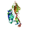
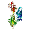
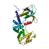
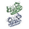
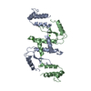
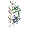
| Title | HUMAN INTERFERON-BETA CRYSTAL STRUCTURE | ||||||||||||
 Components Components | INTERFERON-BETA | ||||||||||||
 Keywords Keywords | INTERFERON / HELICAL CYTOKINE / IMMUNE SYSTEM / CYTOKINE | ||||||||||||
| Function / homology |  Function and homology information Function and homology informationregulation of MHC class I biosynthetic process / negative regulation of Lewy body formation / type I interferon receptor binding / B cell activation involved in immune response / chloramphenicol O-acetyltransferase activity / natural killer cell activation involved in immune response / negative regulation of T cell differentiation / negative regulation of T-helper 2 cell cytokine production / T cell activation involved in immune response / cytokine receptor binding ...regulation of MHC class I biosynthetic process / negative regulation of Lewy body formation / type I interferon receptor binding / B cell activation involved in immune response / chloramphenicol O-acetyltransferase activity / natural killer cell activation involved in immune response / negative regulation of T cell differentiation / negative regulation of T-helper 2 cell cytokine production / T cell activation involved in immune response / cytokine receptor binding / cell surface receptor signaling pathway via STAT / positive regulation of innate immune response / natural killer cell activation / TRAF6 mediated IRF7 activation / negative regulation of viral genome replication / type I interferon-mediated signaling pathway / cellular response to exogenous dsRNA / response to exogenous dsRNA / humoral immune response / B cell proliferation / Regulation of IFNA/IFNB signaling / cell surface receptor signaling pathway via JAK-STAT / cellular response to interferon-beta / positive regulation of autophagy / cytokine activity / positive regulation of apoptotic signaling pathway / neuron cellular homeostasis / cellular response to virus / Evasion by RSV of host interferon responses / response to virus / cytokine-mediated signaling pathway / Interferon alpha/beta signaling / Factors involved in megakaryocyte development and platelet production / TRAF3-dependent IRF activation pathway / Oxidative Stress Induced Senescence / defense response to virus / adaptive immune response / cell surface receptor signaling pathway / innate immune response / SARS-CoV-2 activates/modulates innate and adaptive immune responses / positive regulation of transcription by RNA polymerase II / extracellular space / extracellular region Similarity search - Function | ||||||||||||
| Biological species |  Homo sapiens (human) Homo sapiens (human) | ||||||||||||
| Method |  X-RAY DIFFRACTION / MOLECULAR REPLACEMENT / Resolution: 2.2 Å X-RAY DIFFRACTION / MOLECULAR REPLACEMENT / Resolution: 2.2 Å | ||||||||||||
 Authors Authors | Karpusas, M. / Nolte, M. / Lipscomb, W. | ||||||||||||
 Citation Citation | Journal: Proc.Natl.Acad.Sci.USA / Year: 1997 Title: The crystal structure of human interferon beta at 2.2-A resolution. Authors: Karpusas, M. / Nolte, M. / Benton, C.B. / Meier, W. / Lipscomb, W.N. / Goelz, S. | ||||||||||||
| History |
|
- Structure visualization
| Structure viewer | Molecule: MolmilJmol/JSmol |
|---|
- Downloads & links
-Download
| PDBx/mmCIF format | 1au1.cif.gz | 103.5 KB | Display | PDBx/mmCIF format |
|---|---|---|---|---|
| PDB format | pdb1au1.ent.gz | 80.8 KB | Display | PDB format |
| PDBx/mmJSON format | 1au1.json.gz | Tree view | PDBx/mmJSON format | |
| Others |  Other downloads Other downloads |
-Validation report
| Arichive directory | https://data.pdbj.org/pub/pdb/validation_reports/au/1au1ftp://data.pdbj.org/pub/pdb/validation_reports/au/1au1 | HTTPS FTP |
|---|
-Related structure data
| Similar structure data |
|---|


-Links
 PDBj
PDBj


- Assembly
| Deposited unit | 
| ||||||||
|---|---|---|---|---|---|---|---|---|---|
| 1 |
| ||||||||
| Unit cell |
|
-Components
| #1: Protein | Mass: 20054.080 Da / Num. of mol.: 2 Source method: isolated from a genetically manipulated source Source: (gene. exp.) Homo sapiens (human) / Cell line (production host): CHO / Production host:   Cricetulus griseus (Chinese hamster) / References: UniProt: P01574 Cricetulus griseus (Chinese hamster) / References: UniProt: P01574#2: Polysaccharide | beta-D-glucopyranose-(1-4)-beta-D-glucopyranose-(1-2)-beta-D-glucopyranose-(1-3)-beta-D- ...beta-D-glucopyranose-(1-4)-beta-D-glucopyranose-(1-2)-beta-D-glucopyranose-(1-3)-beta-D-glucopyranose-(1-4)-beta-D-glucopyranose-(1-4)-[alpha-D-quinovopyranose-(1-6)]beta-D-glucopyranose | Source method: isolated from a genetically manipulated source #3: Polysaccharide | alpha-D-quinovopyranose-(1-6)-beta-D-glucopyranose | Source method: isolated from a genetically manipulated source #4: Chemical | ChemComp-ZN / |   Mass: 65.409 Da / Num. of mol.: 1 / Source method: obtained synthetically / Formula: Zn Mass: 65.409 Da / Num. of mol.: 1 / Source method: obtained synthetically / Formula: Zn#5: Water | ChemComp-HOH / |  Mass: 18.015 Da / Num. of mol.: 81 / Source method: isolated from a natural source / Formula: H2O Mass: 18.015 Da / Num. of mol.: 81 / Source method: isolated from a natural source / Formula: H2OHas protein modification | Y | |
|---|
-Experimental details
-Experiment
| Experiment | Method: X-RAY DIFFRACTION / Number of used crystals: 1 |
|---|
- Sample preparation
| Crystal | Density Matthews: 4.83 Å3/Da / Density % sol: 55.43 % | ||||||||||||||||||||||||||||||
|---|---|---|---|---|---|---|---|---|---|---|---|---|---|---|---|---|---|---|---|---|---|---|---|---|---|---|---|---|---|---|---|
| Crystal grow | pH: 7.5 / Details: pH 7.5 | ||||||||||||||||||||||||||||||
| Crystal grow | *PLUS pH: 5 / Method: vapor diffusion, hanging dropDetails: drop solution was mixed with an equal volume of reservoir solution | ||||||||||||||||||||||||||||||
| Components of the solutions | *PLUS
|
-Data collection
| Diffraction | Mean temperature: 108 K |
|---|---|
| Diffraction source | Source: ROTATING ANODE / Type: RIGAKU RUH2R / Wavelength: 1.5418 |
| Detector | Type: RIGAKU / Detector: IMAGE PLATE / Date: Jan 10, 1996 / Details: MIRRORS |
| Radiation | Monochromatic (M) / Laue (L): M / Scattering type: x-ray |
| Radiation wavelength | Wavelength: 1.5418 Å / Relative weight: 1 |
| Reflection | Resolution: 2.2→30 Å / Num. obs: 18405 / % possible obs: 83.1 % / Observed criterion σ(I): 2 / Rmerge(I) obs: 0.0766 |
- Processing
| Software |
| ||||||||||||||||||||||||||||||||||||||||||||||||||||||||||||
|---|---|---|---|---|---|---|---|---|---|---|---|---|---|---|---|---|---|---|---|---|---|---|---|---|---|---|---|---|---|---|---|---|---|---|---|---|---|---|---|---|---|---|---|---|---|---|---|---|---|---|---|---|---|---|---|---|---|---|---|---|---|
| Refinement | Method to determine structure: MOLECULAR REPLACEMENT / Resolution: 2.2→30 Å / Cross valid method: THROUGHOUT / σ(F): 2 Details: RESIDUES 28 - 30 OF MOLECULE B HAVE NOT BEEN MODELED DUE TO WEAK ELECTRON DENSITY.
| ||||||||||||||||||||||||||||||||||||||||||||||||||||||||||||
| Refinement step | Cycle: LAST / Resolution: 2.2→30 Å
| ||||||||||||||||||||||||||||||||||||||||||||||||||||||||||||
| Refine LS restraints |
| ||||||||||||||||||||||||||||||||||||||||||||||||||||||||||||
| Software | *PLUS Name: X-PLOR / Version: 3.81 / Classification: refinement | ||||||||||||||||||||||||||||||||||||||||||||||||||||||||||||
| Refinement | *PLUS | ||||||||||||||||||||||||||||||||||||||||||||||||||||||||||||
| Solvent computation | *PLUS | ||||||||||||||||||||||||||||||||||||||||||||||||||||||||||||
| Displacement parameters | *PLUS |
