 Movie
Movie Controller


+ Open data

- Basic information
| Entry | Database: PDB / ID: 1a3s | ||||||
|---|---|---|---|---|---|---|---|
| Title | HUMAN UBC9 | ||||||
 Components Components | UBC9 | ||||||
 Keywords Keywords | SUMO CONJUGATING ENZYME / UBIQUITIN CONJUGATING ENZYME | ||||||
| Function / homology |  Function and homology information Function and homology informationSUMO conjugating enzyme activity / RING-like zinc finger domain binding / SUMO ligase complex / transferase complex / SUMOylation of nuclear envelope proteins / Negative regulation of activity of TFAP2 (AP-2) family transcription factors / HLH domain binding / SUMO is transferred from E1 to E2 (UBE2I, UBC9) / Vitamin D (calciferol) metabolism / mitotic nuclear membrane reassembly ...SUMO conjugating enzyme activity / RING-like zinc finger domain binding / SUMO ligase complex / transferase complex / SUMOylation of nuclear envelope proteins / Negative regulation of activity of TFAP2 (AP-2) family transcription factors / HLH domain binding / SUMO is transferred from E1 to E2 (UBE2I, UBC9) / Vitamin D (calciferol) metabolism / mitotic nuclear membrane reassembly / synaptonemal complex / small protein activating enzyme binding / SUMOylation of immune response proteins / SUMOylation of DNA methylation proteins / SUMOylation of SUMOylation proteins / Maturation of nucleoprotein / SUMOylation of RNA binding proteins / nuclear export / Transferases; Acyltransferases; Aminoacyltransferases / SUMO transferase activity / Postmitotic nuclear pore complex (NPC) reformation / Maturation of nucleoprotein / SUMOylation of ubiquitinylation proteins / transcription factor binding / SUMOylation of transcription factors / SUMOylation of DNA replication proteins / protein sumoylation / postsynaptic cytosol / nuclear pore / SUMOylation of DNA damage response and repair proteins / SARS-CoV-1 targets host intracellular signalling and regulatory pathways / presynaptic cytosol / Transcriptional and post-translational regulation of MITF-M expression and activity / SUMOylation of transcription cofactors / Meiotic synapsis / SUMOylation of chromatin organization proteins / transcription coregulator binding / Regulation of endogenous retroelements by KRAB-ZFP proteins / SUMOylation of intracellular receptors / chromosome segregation / protein modification process / PKR-mediated signaling / PML body / modulation of chemical synaptic transmission / Schaffer collateral - CA1 synapse / Formation of Incision Complex in GG-NER / nuclear envelope / Recruitment and ATM-mediated phosphorylation of repair and signaling proteins at DNA double strand breaks / Processing of DNA double-strand break ends / ubiquitin-dependent protein catabolic process / positive regulation of canonical NF-kappaB signal transduction / positive regulation of cell migration / cell division / negative regulation of DNA-templated transcription / perinuclear region of cytoplasm / glutamatergic synapse / enzyme binding / negative regulation of transcription by RNA polymerase II / RNA binding / nucleoplasm / ATP binding / nucleus / cytoplasm / cytosol Similarity search - Function | ||||||
| Biological species |  Homo sapiens (human) Homo sapiens (human) | ||||||
| Method |  X-RAY DIFFRACTION / MOLECULAR REPLACEMENT / Resolution: 2.8 Å X-RAY DIFFRACTION / MOLECULAR REPLACEMENT / Resolution: 2.8 Å | ||||||
 Authors Authors | Naismith, J.H. / Giraud, M. | ||||||
 Citation Citation | Journal: Acta Crystallogr.,Sect.D / Year: 1998 Title: Structure of ubiquitin-conjugating enzyme 9 displays significant differences with other ubiquitin-conjugating enzymes which may reflect its specificity for sumo rather than ubiquitin. Authors: Giraud, M.F. / Desterro, J.M. / Naismith, J.H. #1: Journal: To be PublishedTitle: The Structure of Ubch9: A Sumo Conjugating Enzyme Authors: Giraud, M. / Desterro, J.M.P. / Naismith, J.H. | ||||||
| History |
|
- Structure visualization
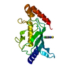
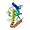
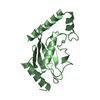
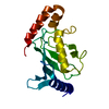
| Structure viewer | Molecule: MolmilJmol/JSmol |
|---|
- Downloads & links
-Download
| PDBx/mmCIF format | 1a3s.cif.gz | 42.4 KB | Display | PDBx/mmCIF format |
|---|---|---|---|---|
| PDB format | pdb1a3s.ent.gz | 30.5 KB | Display | PDB format |
| PDBx/mmJSON format | 1a3s.json.gz | Tree view | PDBx/mmJSON format | |
| Others |  Other downloads Other downloads |
-Validation report
| Arichive directory | https://data.pdbj.org/pub/pdb/validation_reports/a3/1a3sftp://data.pdbj.org/pub/pdb/validation_reports/a3/1a3s | HTTPS FTP |
|---|
-Related structure data
| Related structure data |  1aak S: Starting model for refinement |
|---|---|
| Similar structure data |


-Links
 PDBj
PDBj


- Assembly
| Deposited unit | 
| ||||||||
|---|---|---|---|---|---|---|---|---|---|
| 1 |
| ||||||||
| Unit cell |
|
-Components
| #1: Protein | Mass: 18174.945 Da / Num. of mol.: 1 Source method: isolated from a genetically manipulated source Source: (gene. exp.) Homo sapiens (human) / Description: GST-FUSION / Production host:  |
|---|
-Experimental details
-Experiment
| Experiment | Method: X-RAY DIFFRACTION / Number of used crystals: 1 |
|---|
- Sample preparation
| Crystal | Density Matthews: 3.22 Å3/Da / Density % sol: 61.8 % | ||||||||||||||||||||||||||||||||||||||||
|---|---|---|---|---|---|---|---|---|---|---|---|---|---|---|---|---|---|---|---|---|---|---|---|---|---|---|---|---|---|---|---|---|---|---|---|---|---|---|---|---|---|
| Crystal grow | pH: 7 / Details: pH 7. | ||||||||||||||||||||||||||||||||||||||||
| Crystal grow | *PLUS Temperature: 293.5 K / pH: 8.25 / Method: vapor diffusion, hanging drop | ||||||||||||||||||||||||||||||||||||||||
| Components of the solutions | *PLUS
|
-Data collection
| Diffraction | Mean temperature: 298 K |
|---|---|
| Diffraction source | Source: ROTATING ANODE / Type: ENRAF-NONIUS FR591 / Wavelength: 1.5418 |
| Detector | Type: BRUKER NONIUS / Detector: IMAGE PLATE / Date: Aug 1, 1997 / Details: MIRRORS |
| Radiation | Monochromator: NI FILTER / Monochromatic (M) / Laue (L): M / Scattering type: x-ray |
| Radiation wavelength | Wavelength: 1.5418 Å / Relative weight: 1 |
| Reflection | Highest resolution: 2.8 Å / Num. obs: 5605 / % possible obs: 96 % / Observed criterion σ(I): 0 / Redundancy: 3.1 % / Biso Wilson estimate: 43.8 Å2 / Rmerge(I) obs: 0.1 / Rsym value: 0.1 / Net I/σ(I): 7.2 |
| Reflection shell | Resolution: 2.8→2.9 Å / Redundancy: 2 % / Rmerge(I) obs: 0.19 / Mean I/σ(I) obs: 2.4 / Rsym value: 0.19 / % possible all: 81 |
| Reflection | *PLUS Highest resolution: 2.8 Å / Lowest resolution: 20 Å / Rmerge(I) obs: 0.1 |
| Reflection shell | *PLUS % possible obs: 81.4 % / Num. unique obs: 474 / Rmerge(I) obs: 0.198 |
- Processing
| Software |
| ||||||||||||||||||||||||||||||||||||||||||||||||||||||||||||||||||||||||||||||||
|---|---|---|---|---|---|---|---|---|---|---|---|---|---|---|---|---|---|---|---|---|---|---|---|---|---|---|---|---|---|---|---|---|---|---|---|---|---|---|---|---|---|---|---|---|---|---|---|---|---|---|---|---|---|---|---|---|---|---|---|---|---|---|---|---|---|---|---|---|---|---|---|---|---|---|---|---|---|---|---|---|---|
| Refinement | Method to determine structure: MOLECULAR REPLACEMENT Starting model: PDB ENTRY 1AAK 1aak Rfactor Rfree error: 0.01 / Highest resolution: 2.8 Å / Data cutoff high absF: 9999999999 / Isotropic thermal model: RESTRAINED / Cross valid method: THROUGHOUT / σ(F): 0 Details: ATOMS WITH ZERO OCCUPANCY AR E MODELLED AND WERE NOT LOCATED BY EXPERIMENTAL DENSITY. DATA CUTOFF HIGH (ABS(F)) : 9999999999 DATA CUTOFF LOW (ABS(F)) : 0
| ||||||||||||||||||||||||||||||||||||||||||||||||||||||||||||||||||||||||||||||||
| Solvent computation | Solvent model: DENSITY MODIFICATION / Bsol: 34.51 Å2 / ksol: 0.325 e/Å3 | ||||||||||||||||||||||||||||||||||||||||||||||||||||||||||||||||||||||||||||||||
| Displacement parameters | Biso mean: 42.5 Å2
| ||||||||||||||||||||||||||||||||||||||||||||||||||||||||||||||||||||||||||||||||
| Refine analyze |
| ||||||||||||||||||||||||||||||||||||||||||||||||||||||||||||||||||||||||||||||||
| Refinement step | Cycle: LAST / Highest resolution: 2.8 Å
| ||||||||||||||||||||||||||||||||||||||||||||||||||||||||||||||||||||||||||||||||
| Refine LS restraints |
| ||||||||||||||||||||||||||||||||||||||||||||||||||||||||||||||||||||||||||||||||
| LS refinement shell | Resolution: 2.8→2.9 Å / Rfactor Rfree error: 0.07 / Total num. of bins used: 10
| ||||||||||||||||||||||||||||||||||||||||||||||||||||||||||||||||||||||||||||||||
| Xplor file |
| ||||||||||||||||||||||||||||||||||||||||||||||||||||||||||||||||||||||||||||||||
| Software | *PLUS Name: CNS / Version: 0.2.0 / Classification: refinement | ||||||||||||||||||||||||||||||||||||||||||||||||||||||||||||||||||||||||||||||||
| Refinement | *PLUS Lowest resolution: 26 Å | ||||||||||||||||||||||||||||||||||||||||||||||||||||||||||||||||||||||||||||||||
| Solvent computation | *PLUS | ||||||||||||||||||||||||||||||||||||||||||||||||||||||||||||||||||||||||||||||||
| Displacement parameters | *PLUS | ||||||||||||||||||||||||||||||||||||||||||||||||||||||||||||||||||||||||||||||||
| Refine LS restraints | *PLUS
|
