 Movie
Movie Controller


+ Open data

- Basic information
| Entry | Database: PDB / ID: 1a21 | ||||||
|---|---|---|---|---|---|---|---|
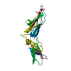
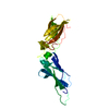
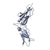
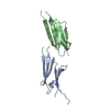
| Title | TISSUE FACTOR (TF) FROM RABBIT | ||||||
 Components Components | TISSUE FACTOR | ||||||
 Keywords Keywords | GLYCOPROTEIN / BLOOD COAGULATION FACTOR / FVIIA ACTIVATION / CYTOKINE RECEPTOR SUPERFAMILY / EXTRACELLULAR DOMAIN | ||||||
| Function / homology |  Function and homology information Function and homology informationactivation of plasma proteins involved in acute inflammatory response / positive regulation of platelet-derived growth factor receptor signaling pathway / cytokine receptor activity / positive regulation of endothelial cell apoptotic process / positive regulation of TOR signaling / positive regulation of endothelial cell proliferation / phospholipid binding / extracellular matrix / positive regulation of angiogenesis / blood coagulation ...activation of plasma proteins involved in acute inflammatory response / positive regulation of platelet-derived growth factor receptor signaling pathway / cytokine receptor activity / positive regulation of endothelial cell apoptotic process / positive regulation of TOR signaling / positive regulation of endothelial cell proliferation / phospholipid binding / extracellular matrix / positive regulation of angiogenesis / blood coagulation / cell surface / extracellular space / plasma membrane Similarity search - Function | ||||||
| Biological species |  | ||||||
| Method |  X-RAY DIFFRACTION / SYNCHROTRON / MOLECULAR REPLACEMENT / Resolution: 2.35 Å X-RAY DIFFRACTION / SYNCHROTRON / MOLECULAR REPLACEMENT / Resolution: 2.35 Å | ||||||
 Authors Authors | Muller, Y.A. / De Vos, A.M. | ||||||
 Citation Citation | Journal: Protein Sci. / Year: 1998 Title: Hinge bending within the cytokine receptor superfamily revealed by the 2.4 A crystal structure of the extracellular domain of rabbit tissue factor. Authors: Muller, Y.A. / Kelley, R.F. / de Vos, A.M. | ||||||
| History |
|
- Structure visualization
| Structure viewer | Molecule: MolmilJmol/JSmol |
|---|
- Downloads & links
-Download
| PDBx/mmCIF format | 1a21.cif.gz | 95.5 KB | Display | PDBx/mmCIF format |
|---|---|---|---|---|
| PDB format | pdb1a21.ent.gz | 72.7 KB | Display | PDB format |
| PDBx/mmJSON format | 1a21.json.gz | Tree view | PDBx/mmJSON format | |
| Others |  Other downloads Other downloads |
-Validation report
| Arichive directory | https://data.pdbj.org/pub/pdb/validation_reports/a2/1a21ftp://data.pdbj.org/pub/pdb/validation_reports/a2/1a21 | HTTPS FTP |
|---|
-Related structure data
| Related structure data | 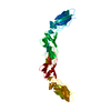 2hftS S: Starting model for refinement |
|---|---|
| Similar structure data |


-Links
 PDBj
PDBj

- Assembly
| Deposited unit | 
| ||||||||||||
|---|---|---|---|---|---|---|---|---|---|---|---|---|---|
| 1 | 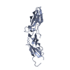
| ||||||||||||
| 2 | 
| ||||||||||||
| Unit cell |
| ||||||||||||
| Noncrystallographic symmetry (NCS) | NCS oper:
|
-Components
| #1: Protein | Mass: 25106.051 Da / Num. of mol.: 2 / Fragment: EXTRACELLULAR DOMAIN, RESIDUES 1 - 219 Source method: isolated from a genetically manipulated source Source: (gene. exp.)  #2: Water | ChemComp-HOH / |  Mass: 18.015 Da / Num. of mol.: 231 / Source method: isolated from a natural source / Formula: H2O Mass: 18.015 Da / Num. of mol.: 231 / Source method: isolated from a natural source / Formula: H2OHas protein modification | Y | |
|---|
-Experimental details
-Experiment
| Experiment | Method: X-RAY DIFFRACTION / Number of used crystals: 1 |
|---|
- Sample preparation
| Crystal | Density Matthews: 2.7 Å3/Da / Density % sol: 54 % | ||||||||||||||||||||||||||||||||||||||||||||||||||||||||||||
|---|---|---|---|---|---|---|---|---|---|---|---|---|---|---|---|---|---|---|---|---|---|---|---|---|---|---|---|---|---|---|---|---|---|---|---|---|---|---|---|---|---|---|---|---|---|---|---|---|---|---|---|---|---|---|---|---|---|---|---|---|---|
| Crystal grow | Method: vapor diffusion, sitting drop / pH: 8 Details: SITTING DROP, 80 UL OF PROTEIN SOLUTION (10 MG/ML PROTEIN IN 50 MM TRIS*HCL,PH:8.0, 50 MM NACL, 75 MM LI2SO4, 12.5 % PEG 3350) EQUILIBRATED AGAINST 30 ML RESERVOIR SOLUTION (50 MM TRIS*HCL, ...Details: SITTING DROP, 80 UL OF PROTEIN SOLUTION (10 MG/ML PROTEIN IN 50 MM TRIS*HCL,PH:8.0, 50 MM NACL, 75 MM LI2SO4, 12.5 % PEG 3350) EQUILIBRATED AGAINST 30 ML RESERVOIR SOLUTION (50 MM TRIS*HCL, PH:8.0, 50 MM NACL, 150 MM LI2SO4, 25 % PEG3350), vapor diffusion - sitting drop | ||||||||||||||||||||||||||||||||||||||||||||||||||||||||||||
| Crystal grow | *PLUS Method: vapor diffusion, sitting drop | ||||||||||||||||||||||||||||||||||||||||||||||||||||||||||||
| Components of the solutions | *PLUS
|
-Data collection
| Diffraction | Mean temperature: 100 K |
|---|---|
| Diffraction source | Source: SYNCHROTRON / Site: CHESS  / Beamline: F1 / Wavelength: 0.918 / Beamline: F1 / Wavelength: 0.918 |
| Detector | Type: PRINCETON 2K / Detector: CCD / Date: Mar 1, 1996 |
| Radiation | Monochromatic (M) / Laue (L): M / Scattering type: x-ray |
| Radiation wavelength | Wavelength: 0.918 Å / Relative weight: 1 |
| Reflection | Resolution: 2.35→60 Å / Num. obs: 20678 / % possible obs: 96.4 % / Redundancy: 3 % / Biso Wilson estimate: 35.1 Å2 / Rmerge(I) obs: 0.075 |
| Reflection shell | Resolution: 2.35→2.4 Å / Redundancy: 2 % / Rsym value: 0.07 / % possible all: 90.6 |
| Reflection shell | *PLUS % possible obs: 90.6 % / Rmerge(I) obs: 0.07 |
- Processing
| Software |
| ||||||||||||||||||||||||||||||||||||||||||||||||||||||||||||||||||||||||||||||||
|---|---|---|---|---|---|---|---|---|---|---|---|---|---|---|---|---|---|---|---|---|---|---|---|---|---|---|---|---|---|---|---|---|---|---|---|---|---|---|---|---|---|---|---|---|---|---|---|---|---|---|---|---|---|---|---|---|---|---|---|---|---|---|---|---|---|---|---|---|---|---|---|---|---|---|---|---|---|---|---|---|---|
| Refinement | Method to determine structure: MOLECULAR REPLACEMENT Starting model: PDB ENTRY 2HFT Resolution: 2.35→10 Å / Rfactor Rfree error: 0.006 / Data cutoff high absF: 100000 / Data cutoff low absF: 0.1 / Isotropic thermal model: RESTRAINED / Cross valid method: THROUGHOUT Details: A BULK SOLVENT CORRECTION CALCULATED WITH PROGRAM X-PLOR WAS USED THROUGHOUT REFINEMENT.
| ||||||||||||||||||||||||||||||||||||||||||||||||||||||||||||||||||||||||||||||||
| Displacement parameters | Biso mean: 34.9 Å2
| ||||||||||||||||||||||||||||||||||||||||||||||||||||||||||||||||||||||||||||||||
| Refine analyze | Luzzati coordinate error obs: 0.25 Å / Luzzati d res low obs: 10 Å / Luzzati sigma a obs: 0.25 Å | ||||||||||||||||||||||||||||||||||||||||||||||||||||||||||||||||||||||||||||||||
| Refinement step | Cycle: LAST / Resolution: 2.35→10 Å
| ||||||||||||||||||||||||||||||||||||||||||||||||||||||||||||||||||||||||||||||||
| Refine LS restraints |
| ||||||||||||||||||||||||||||||||||||||||||||||||||||||||||||||||||||||||||||||||
| LS refinement shell | Resolution: 2.35→2.4 Å / Rfactor Rfree error: 0.031 / Total num. of bins used: 15
| ||||||||||||||||||||||||||||||||||||||||||||||||||||||||||||||||||||||||||||||||
| Xplor file |
| ||||||||||||||||||||||||||||||||||||||||||||||||||||||||||||||||||||||||||||||||
| Software | *PLUS Name: X-PLOR / Version: 3.851 / Classification: refinement | ||||||||||||||||||||||||||||||||||||||||||||||||||||||||||||||||||||||||||||||||
| Refinement | *PLUS | ||||||||||||||||||||||||||||||||||||||||||||||||||||||||||||||||||||||||||||||||
| Solvent computation | *PLUS | ||||||||||||||||||||||||||||||||||||||||||||||||||||||||||||||||||||||||||||||||
| Displacement parameters | *PLUS | ||||||||||||||||||||||||||||||||||||||||||||||||||||||||||||||||||||||||||||||||
| Refine LS restraints | *PLUS
| ||||||||||||||||||||||||||||||||||||||||||||||||||||||||||||||||||||||||||||||||
| LS refinement shell | *PLUS Rfactor obs: 0.299 |
