 Movie
Movie Controller

[English] 日本語
 Yorodumi
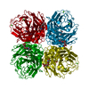
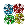
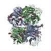
- PDB-6hp0: Complex of Neuraminidase from H1N1 Influenza Virus in Complex wit... -
+ Open data

- Basic information
| Entry | Database: PDB / ID: 6hp0 | |||||||||
|---|---|---|---|---|---|---|---|---|---|---|
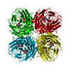
| Title | Complex of Neuraminidase from H1N1 Influenza Virus in Complex with Oseltamivir Triazol Derivative | |||||||||
 Components Components | Neuraminidase | |||||||||
 Keywords Keywords | VIRAL PROTEIN / neuraminidase / influenza / complex / inhibitor | |||||||||
| Function / homology |  Function and homology information Function and homology informationexo-alpha-(2->3)-sialidase activity / exo-alpha-(2->6)-sialidase activity / exo-alpha-(2->8)-sialidase activity / exo-alpha-sialidase / viral budding from plasma membrane / carbohydrate metabolic process / host cell plasma membrane / virion membrane / membrane / metal ion bindingSimilarity search - Function | |||||||||
| Biological species |   Influenza A virus Influenza A virus | |||||||||
| Method | X-RAY DIFFRACTION / SYNCHROTRON / MOLECULAR REPLACEMENT / Resolution: 1.88 Å | |||||||||
 Authors Authors | Pachl, P. / Pokorna, J. | |||||||||
| Funding support |  Czech Republic, 1items Czech Republic, 1items
| |||||||||
 Citation Citation | Journal: Bioorg.Med.Chem. / Year: 2019 Title: Investigation of flexibility of neuraminidase 150-loop using tamiflu derivatives in influenza A viruses H1N1 and H5N1. Authors: Zima, V. / Albinana, C.B. / Rojikova, K. / Pokorna, J. / Pachl, P. / Rezacova, P. / Hudlicky, J. / Navratil, V. / Majer, P. / Konvalinka, J. / Kozisek, M. / Machara, A. | |||||||||
| History |
|
- Structure visualization
| Structure viewer | Molecule: MolmilJmol/JSmol |
|---|
- Downloads & links
-Download
| PDBx/mmCIF format | 6hp0.cif.gz | 355.6 KB | Display | PDBx/mmCIF format |
|---|---|---|---|---|
| PDB format | pdb6hp0.ent.gz | 285.8 KB | Display | PDB format |
| PDBx/mmJSON format | 6hp0.json.gz | Tree view | PDBx/mmJSON format | |
| Others |  Other downloads Other downloads |
-Validation report
| Arichive directory | https://data.pdbj.org/pub/pdb/validation_reports/hp/6hp0ftp://data.pdbj.org/pub/pdb/validation_reports/hp/6hp0 | HTTPS FTP |
|---|
-Related structure data
| Related structure data | 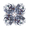 6g02S S: Starting model for refinement |
|---|---|
| Similar structure data |


-Links
 PDBj
PDBj

- Assembly
| Deposited unit | 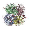
| ||||||||
|---|---|---|---|---|---|---|---|---|---|
| 1 |
| ||||||||
| Unit cell |
|
-Components
-Protein , 1 types, 4 molecules ABCD
| #1: Protein | Mass: 42727.629 Da / Num. of mol.: 4 Source method: isolated from a genetically manipulated source Source: (gene. exp.) Influenza A virus (A/Texas/17/2009(H1N1))Gene: NA / Production host:  Drosophila melanogaster (fruit fly) / References: UniProt: C6KP13, exo-alpha-sialidase Drosophila melanogaster (fruit fly) / References: UniProt: C6KP13, exo-alpha-sialidase |
|---|
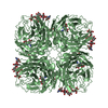
-Sugars , 4 types, 12 molecules 
| #2: Polysaccharide | / Mass: 570.542 Da / Num. of mol.: 3 Source method: isolated from a genetically manipulated source #3: Polysaccharide | / Mass: 367.349 Da / Num. of mol.: 2Source method: isolated from a genetically manipulated source #4: Polysaccharide | alpha-D-mannopyranose-(1-3)-[alpha-D-mannopyranose-(1-6)]beta-D-mannopyranose-(1-4)-2-acetamido-2- ...alpha-D-mannopyranose-(1-3)-[alpha-D-mannopyranose-(1-6)]beta-D-mannopyranose-(1-4)-2-acetamido-2-deoxy-beta-D-glucopyranose-(1-4)-[alpha-L-fucopyranose-(1-6)]2-acetamido-2-deoxy-beta-D-glucopyranose | / Mass: 1056.964 Da / Num. of mol.: 1Source method: isolated from a genetically manipulated source #7: Sugar | ChemComp-NAG / N-Acetylglucosamine Type: D-saccharide, beta linking / Mass: 221.208 Da / Num. of mol.: 6 Type: D-saccharide, beta linking / Mass: 221.208 Da / Num. of mol.: 6Source method: isolated from a genetically manipulated source Formula: C8H15NO6 |
|---|
-Non-polymers , 5 types, 1441 molecules 
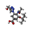


| #5: Chemical | ChemComp-CA /  Mass: 40.078 Da / Num. of mol.: 8 / Source method: obtained synthetically / Formula: Ca Mass: 40.078 Da / Num. of mol.: 8 / Source method: obtained synthetically / Formula: Ca#6: Chemical | ChemComp-GJT / (  Mass: 366.412 Da / Num. of mol.: 4 / Source method: obtained synthetically / Formula: C17H26N4O5 Mass: 366.412 Da / Num. of mol.: 4 / Source method: obtained synthetically / Formula: C17H26N4O5#8: Chemical | ChemComp-EDO / Ethylene glycol Mass: 62.068 Da / Num. of mol.: 29 / Source method: obtained synthetically / Formula: C2H6O2 Mass: 62.068 Da / Num. of mol.: 29 / Source method: obtained synthetically / Formula: C2H6O2#9: Chemical | ChemComp-PGE / | Polyethylene glycol Mass: 150.173 Da / Num. of mol.: 1 / Source method: obtained synthetically / Formula: C6H14O4 Mass: 150.173 Da / Num. of mol.: 1 / Source method: obtained synthetically / Formula: C6H14O4#10: Water | ChemComp-HOH / | WaterMass: 18.015 Da / Num. of mol.: 1399 / Source method: isolated from a natural source / Formula: H2O |
|---|
-Experimental details
-Experiment
| Experiment | Method: X-RAY DIFFRACTION / Number of used crystals: 1 |
|---|
- Sample preparation
| Crystal | Density Matthews: 3.08 Å3/Da / Density % sol: 60.07 % |
|---|---|
| Crystal grow | Temperature: 291 K / Method: vapor diffusion, hanging drop / pH: 8 / Details: 0.1 M HEPES pH 7.5, 5% PEG 8000 |
-Data collection
| Diffraction | Mean temperature: 100 K |
|---|---|
| Diffraction source | Source: SYNCHROTRON / Site: BESSY  / Beamline: 14.1 / Wavelength: 0.9184 Å / Beamline: 14.1 / Wavelength: 0.9184 Å |
| Detector | Type: DECTRIS PILATUS 6M / Detector: PIXEL / Date: Sep 29, 2015 |
| Radiation | Protocol: SINGLE WAVELENGTH / Monochromatic (M) / Laue (L): M / Scattering type: x-ray |
| Radiation wavelength | Wavelength: 0.9184 Å / Relative weight: 1 |
| Reflection | Resolution: 1.88→48.43 Å / Num. obs: 162570 / % possible obs: 97.6 % / Redundancy: 2.1 % / Biso Wilson estimate: 26.8 Å2 / CC1/2: 0.996 / Rrim(I) all: 0.096 / Net I/σ(I): 9.24 |
| Reflection shell | Resolution: 1.88→2 Å / Redundancy: 2.3 % / Mean I/σ(I) obs: 1.96 / Num. unique obs: 26059 / CC1/2: 0.723 / Rrim(I) all: 0.558 / % possible all: 97 |
- Processing
| Software |
| ||||||||||||||||||||||||||||||||||||||||||||||||||||||||||||||||||||||||||||||||||||||||||||||||||||||||||||||||||||||||||||||||||||||||||||||||||||||||||||||||||||||||||||||||||||||
|---|---|---|---|---|---|---|---|---|---|---|---|---|---|---|---|---|---|---|---|---|---|---|---|---|---|---|---|---|---|---|---|---|---|---|---|---|---|---|---|---|---|---|---|---|---|---|---|---|---|---|---|---|---|---|---|---|---|---|---|---|---|---|---|---|---|---|---|---|---|---|---|---|---|---|---|---|---|---|---|---|---|---|---|---|---|---|---|---|---|---|---|---|---|---|---|---|---|---|---|---|---|---|---|---|---|---|---|---|---|---|---|---|---|---|---|---|---|---|---|---|---|---|---|---|---|---|---|---|---|---|---|---|---|---|---|---|---|---|---|---|---|---|---|---|---|---|---|---|---|---|---|---|---|---|---|---|---|---|---|---|---|---|---|---|---|---|---|---|---|---|---|---|---|---|---|---|---|---|---|---|---|---|---|
| Refinement | Method to determine structure: MOLECULAR REPLACEMENT Starting model: 6G02 Resolution: 1.88→48.34 Å / Cor.coef. Fo:Fc: 0.954 / Cor.coef. Fo:Fc free: 0.936 / SU B: 3.816 / SU ML: 0.108 / Cross valid method: THROUGHOUT / ESU R: 0.129 / ESU R Free: 0.125 / Stereochemistry target values: MAXIMUM LIKELIHOOD / Details: HYDROGENS HAVE BEEN ADDED IN THE RIDING POSITIONS
| ||||||||||||||||||||||||||||||||||||||||||||||||||||||||||||||||||||||||||||||||||||||||||||||||||||||||||||||||||||||||||||||||||||||||||||||||||||||||||||||||||||||||||||||||||||||
| Solvent computation | Ion probe radii: 0.8 Å / Shrinkage radii: 0.8 Å / VDW probe radii: 1.2 Å / Solvent model: MASK | ||||||||||||||||||||||||||||||||||||||||||||||||||||||||||||||||||||||||||||||||||||||||||||||||||||||||||||||||||||||||||||||||||||||||||||||||||||||||||||||||||||||||||||||||||||||
| Displacement parameters | Biso mean: 21.873 Å2
| ||||||||||||||||||||||||||||||||||||||||||||||||||||||||||||||||||||||||||||||||||||||||||||||||||||||||||||||||||||||||||||||||||||||||||||||||||||||||||||||||||||||||||||||||||||||
| Refinement step | Cycle: 1 / Resolution: 1.88→48.34 Å
| ||||||||||||||||||||||||||||||||||||||||||||||||||||||||||||||||||||||||||||||||||||||||||||||||||||||||||||||||||||||||||||||||||||||||||||||||||||||||||||||||||||||||||||||||||||||
| Refine LS restraints |
|