 Movie
Movie Controller

[English] 日本語
 Yorodumi
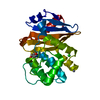
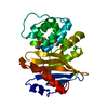
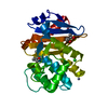
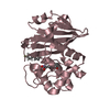
- PDB-5fq1: Structure of the cytoplasmic PAS domain of the Geobacillus thermo... -
+ Open data

- Basic information
| Entry | Database: PDB / ID: 5fq1 | ||||||
|---|---|---|---|---|---|---|---|
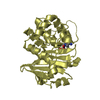
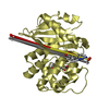
| Title | Structure of the cytoplasmic PAS domain of the Geobacillus thermodenitrificans histidine kinase CitA | ||||||
 Components Components | HISTIDINE KINASE | ||||||
 Keywords Keywords | TRANSFERASE / PAS DOMAIN / CITA / TRANSMEMBRANE SIGNALING | ||||||
| Function / homology |  Function and homology informationhistidine kinase / phosphorelay sensor kinase activity / membrane => GO:0016020 / ATP binding / plasma membrane Function and homology informationhistidine kinase / phosphorelay sensor kinase activity / membrane => GO:0016020 / ATP binding / plasma membraneSimilarity search - Function | ||||||
| Biological species |  GEOBACILLUS THERMODENITRIFICANS (bacteria) GEOBACILLUS THERMODENITRIFICANS (bacteria) | ||||||
| Method | X-RAY DIFFRACTION / SYNCHROTRON / MAD / Resolution: 1.76 Å | ||||||
 Authors Authors | Schomburg, B. / Giller, K. / Becker, S. | ||||||
 Citation Citation | Journal: Nat. Methods / Year: 2017 Title: Cryogenic optical localization provides 3D protein structure data with Angstrom resolution. Authors: Weisenburger, S. / Boening, D. / Schomburg, B. / Giller, K. / Becker, S. / Griesinger, C. / Sandoghdar, V. | ||||||
| History |
|
- Structure visualization
| Structure viewer | Molecule: MolmilJmol/JSmol |
|---|
- Downloads & links
-Download
| PDBx/mmCIF format | 5fq1.cif.gz | 53.6 KB | Display | PDBx/mmCIF format |
|---|---|---|---|---|
| PDB format | pdb5fq1.ent.gz | 42.6 KB | Display | PDB format |
| PDBx/mmJSON format | 5fq1.json.gz | Tree view | PDBx/mmJSON format | |
| Others |  Other downloads Other downloads |
-Validation report
| Arichive directory | https://data.pdbj.org/pub/pdb/validation_reports/fq/5fq1ftp://data.pdbj.org/pub/pdb/validation_reports/fq/5fq1 | HTTPS FTP |
|---|
-Related structure data
| Similar structure data |
|---|


-Links
 PDBj
PDBj


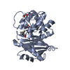
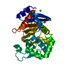
- Assembly
| Deposited unit | 
| ||||||||
|---|---|---|---|---|---|---|---|---|---|
| 1 |
| ||||||||
| Unit cell |
|
-Components
| #1: Protein | / CITA Mass: 12444.914 Da / Num. of mol.: 2 / Fragment: CYTOPLASMIC PAS DOMAIN, UNP RESIDUES 200-309 Source method: isolated from a genetically manipulated source Source: (gene. exp.) GEOBACILLUS THERMODENITRIFICANS (bacteria)Description: DSM466 / Plasmid: PET28A / Production host: ESCHERICHIA COLI (E. coli) / Strain (production host): BL21(DE3) / References: UniProt: A4IPE6, histidine kinase#2: Chemical | ChemComp-GOL / Glycerol  Mass: 92.094 Da / Num. of mol.: 4 / Source method: obtained synthetically / Formula: C3H8O3 Mass: 92.094 Da / Num. of mol.: 4 / Source method: obtained synthetically / Formula: C3H8O3#3: Chemical | Phosphate  Mass: 94.971 Da / Num. of mol.: 3 / Source method: obtained synthetically / Formula: PO4 Mass: 94.971 Da / Num. of mol.: 3 / Source method: obtained synthetically / Formula: PO4#4: Water | ChemComp-HOH / | Water Mass: 18.015 Da / Num. of mol.: 51 / Source method: isolated from a natural source / Formula: H2O Mass: 18.015 Da / Num. of mol.: 51 / Source method: isolated from a natural source / Formula: H2OSequence details | N-TERMINAL GS IS DUE TO CLONING | |
|---|
-Experimental details
-Experiment
| Experiment | Method: X-RAY DIFFRACTION / Number of used crystals: 1 |
|---|
- Sample preparation
| Crystal | Density Matthews: 1.96 Å3/Da / Density % sol: 37 % / Description: NONE |
|---|---|
| Crystal grow | pH: 4.7 Details: 0.4 M K2HPO4, 1.6 M NAH2PO4, 0.1 M PHOSPHATE-CITRATE, PH 4.7 |
-Data collection
| Diffraction | Mean temperature: 100 K | |||||||||||||||
|---|---|---|---|---|---|---|---|---|---|---|---|---|---|---|---|---|
| Diffraction source | Source: SYNCHROTRON / Site: SLS  / Beamline: X10SA / Wavelength: 0.97929 / Beamline: X10SA / Wavelength: 0.97929 | |||||||||||||||
| Detector | Type: DECTRIS PILATUS 6M / Detector: PIXEL / Date: Sep 19, 2012 | |||||||||||||||
| Radiation | Protocol: MAD / Monochromatic (M) / Laue (L): M / Scattering type: x-ray | |||||||||||||||
| Radiation wavelength | Wavelength: 0.97929 Å / Relative weight: 1 | |||||||||||||||
| Reflection twin |
| |||||||||||||||
| Reflection | Resolution: 1.78→37.54 Å / Num. obs: 18999 / % possible obs: 99.3 % / Observed criterion σ(I): 2 / Redundancy: 6.34 % / Rmerge(I) obs: 0.09 / Net I/σ(I): 10.78 | |||||||||||||||
| Reflection shell | Resolution: 1.78→1.88 Å / Redundancy: 6.14 % / Rmerge(I) obs: 0.51 / Mean I/σ(I) obs: 2.4 / % possible all: 96.5 |
- Processing
| Software |
| ||||||||||||||||||||||||||||||||||||||||||||||||||||||||||||||||||||||||||||||||||||||||||||||||||||||||||||||||||||||||||||||||||||||||||||||||||||||||||||||||||||||||||||||||||||||
|---|---|---|---|---|---|---|---|---|---|---|---|---|---|---|---|---|---|---|---|---|---|---|---|---|---|---|---|---|---|---|---|---|---|---|---|---|---|---|---|---|---|---|---|---|---|---|---|---|---|---|---|---|---|---|---|---|---|---|---|---|---|---|---|---|---|---|---|---|---|---|---|---|---|---|---|---|---|---|---|---|---|---|---|---|---|---|---|---|---|---|---|---|---|---|---|---|---|---|---|---|---|---|---|---|---|---|---|---|---|---|---|---|---|---|---|---|---|---|---|---|---|---|---|---|---|---|---|---|---|---|---|---|---|---|---|---|---|---|---|---|---|---|---|---|---|---|---|---|---|---|---|---|---|---|---|---|---|---|---|---|---|---|---|---|---|---|---|---|---|---|---|---|---|---|---|---|---|---|---|---|---|---|---|
| Refinement | Method to determine structure: MAD Starting model: NONE Resolution: 1.76→37.54 Å / Cor.coef. Fo:Fc: 0.963 / Cor.coef. Fo:Fc free: 0.949 / SU B: 2.658 / SU ML: 0.086 / Cross valid method: THROUGHOUT / ESU R: 0.029 / ESU R Free: 0.026 / Stereochemistry target values: MAXIMUM LIKELIHOOD / Details: HYDROGENS HAVE BEEN ADDED IN THE RIDING POSITIONS.
| ||||||||||||||||||||||||||||||||||||||||||||||||||||||||||||||||||||||||||||||||||||||||||||||||||||||||||||||||||||||||||||||||||||||||||||||||||||||||||||||||||||||||||||||||||||||
| Solvent computation | Ion probe radii: 0.8 Å / Shrinkage radii: 0.8 Å / VDW probe radii: 1.2 Å / Solvent model: MASK | ||||||||||||||||||||||||||||||||||||||||||||||||||||||||||||||||||||||||||||||||||||||||||||||||||||||||||||||||||||||||||||||||||||||||||||||||||||||||||||||||||||||||||||||||||||||
| Displacement parameters | Biso mean: 29.382 Å2
| ||||||||||||||||||||||||||||||||||||||||||||||||||||||||||||||||||||||||||||||||||||||||||||||||||||||||||||||||||||||||||||||||||||||||||||||||||||||||||||||||||||||||||||||||||||||
| Refinement step | Cycle: LAST / Resolution: 1.76→37.54 Å
| ||||||||||||||||||||||||||||||||||||||||||||||||||||||||||||||||||||||||||||||||||||||||||||||||||||||||||||||||||||||||||||||||||||||||||||||||||||||||||||||||||||||||||||||||||||||
| Refine LS restraints |
|