 Movie
Movie Controller

[English] 日本語
 Yorodumi
- PDB-5b7z: Crystal Structure of Hyperthermophilic Thermotoga maritima L-Keto... -
+ Open data

- Basic information
| Entry | Database: PDB / ID: 5b7z | |||||||||
|---|---|---|---|---|---|---|---|---|---|---|
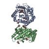
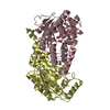
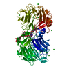
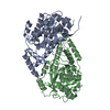
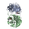
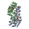
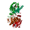
| Title | Crystal Structure of Hyperthermophilic Thermotoga maritima L-Ketose-3-Epimerase with Ni2+ | |||||||||
 Components Components | Uncharacterized protein TM_0416 | |||||||||
 Keywords Keywords | ISOMERASE / epimerase / Ni2+ / hyperthermophilic / eubacterium | |||||||||
| Function / homology |  Function and homology information Function and homology informationIsomerases; Intramolecular oxidoreductases; Interconverting aldoses and ketoses, and related compounds / Isomerases; Racemases and epimerases; Acting on carbohydrates and derivatives / inositol metabolic process / isomerase activity / metal ion binding Similarity search - Function | |||||||||
| Biological species |   Thermotoga maritima MSB8 (bacteria) Thermotoga maritima MSB8 (bacteria) | |||||||||
| Method |  X-RAY DIFFRACTION / SYNCHROTRON / MOLECULAR REPLACEMENT / Resolution: 1.5 Å X-RAY DIFFRACTION / SYNCHROTRON / MOLECULAR REPLACEMENT / Resolution: 1.5 Å | |||||||||
 Authors Authors | Cao, T.P. / Shin, S.M. / Lee, D.W. / Lee, S.H. | |||||||||
| Funding support |  Korea, Republic Of, 2items Korea, Republic Of, 2items
| |||||||||
 Citation Citation | Journal: Appl. Environ. Microbiol. / Year: 2017 Title: TM0416, a Hyperthermophilic Promiscuous Nonphosphorylated Sugar Isomerase, Catalyzes Various C5and C6Epimerization Reactions Authors: Shin, S.M. / Cao, T.P. / Choi, J.M. / Kim, S.B. / Lee, S.J. / Lee, S.H. / Lee, D.W. | |||||||||
| History |
|
- Structure visualization
| Structure viewer | Molecule: MolmilJmol/JSmol |
|---|
- Downloads & links
-Download
| PDBx/mmCIF format | 5b7z.cif.gz | 141.4 KB | Display | PDBx/mmCIF format |
|---|---|---|---|---|
| PDB format | pdb5b7z.ent.gz | 106.6 KB | Display | PDB format |
| PDBx/mmJSON format | 5b7z.json.gz | Tree view | PDBx/mmJSON format | |
| Others |  Other downloads Other downloads |
-Validation report
| Arichive directory | https://data.pdbj.org/pub/pdb/validation_reports/b7/5b7zftp://data.pdbj.org/pub/pdb/validation_reports/b7/5b7z | HTTPS FTP |
|---|
-Related structure data
| Related structure data |  5b7ySC  5b80C 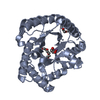 5h1wC 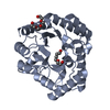 5h6hC S: Starting model for refinement C: citing same article ( |
|---|---|
| Similar structure data |


-Links
 PDBj
PDBj
- Assembly
| Deposited unit | 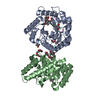
| ||||||||
|---|---|---|---|---|---|---|---|---|---|
| 1 |
| ||||||||
| Unit cell |
|
-Components
| #1: Protein | Mass: 32677.482 Da / Num. of mol.: 2 Source method: isolated from a genetically manipulated source Source: (gene. exp.) Thermotoga maritima MSB8 (bacteria) / Strain: MSB8 / Gene: TM_0416 / Plasmid: pET15b / Production host: #2: Chemical | ChemComp-MPD / (   Mass: 118.174 Da / Num. of mol.: 5 / Source method: obtained synthetically / Formula: C6H14O2 / Comment: precipitant*YM Mass: 118.174 Da / Num. of mol.: 5 / Source method: obtained synthetically / Formula: C6H14O2 / Comment: precipitant*YM#3: Chemical |   Mass: 58.693 Da / Num. of mol.: 2 / Source method: obtained synthetically / Formula: Ni Mass: 58.693 Da / Num. of mol.: 2 / Source method: obtained synthetically / Formula: Ni#4: Chemical | ChemComp-1PG /   Mass: 252.305 Da / Num. of mol.: 4 / Source method: obtained synthetically / Formula: C11H24O6 Mass: 252.305 Da / Num. of mol.: 4 / Source method: obtained synthetically / Formula: C11H24O6#5: Water | ChemComp-HOH / |  Mass: 18.015 Da / Num. of mol.: 670 / Source method: isolated from a natural source / Formula: H2O Mass: 18.015 Da / Num. of mol.: 670 / Source method: isolated from a natural source / Formula: H2O |
|---|
-Experimental details
-Experiment
| Experiment | Method: X-RAY DIFFRACTION / Number of used crystals: 1 |
|---|
- Sample preparation
| Crystal | Density Matthews: 2.31 Å3/Da / Density % sol: 46.9 % |
|---|---|
| Crystal grow | Temperature: 293 K / Method: vapor diffusion, hanging drop / pH: 6 / Details: 100mM MES, 5%(w/w) PEG1000, 20%(v/v) PEG200 |
-Data collection
| Diffraction | Mean temperature: 100 K |
|---|---|
| Diffraction source | Source: SYNCHROTRON / Site: PAL/PLS / Beamline: 5C (4A) / Wavelength: 0.9796 Å |
| Detector | Type: ADSC QUANTUM 315r / Detector: CCD / Date: Jun 3, 2016 |
| Radiation | Monochromator: DCM Si (111) Crystal / Protocol: SINGLE WAVELENGTH / Monochromatic (M) / Laue (L): M / Scattering type: x-ray |
| Radiation wavelength | Wavelength: 0.9796 Å / Relative weight: 1 |
| Reflection | Resolution: 1.5→50 Å / Num. obs: 90302 / % possible obs: 96 % / Redundancy: 3.5 % / Net I/σ(I): 13.27 |
| Reflection shell | Resolution: 1.5→1.53 Å / Redundancy: 3.3 % / Mean I/σ(I) obs: 3.13 / % possible all: 92.8 |
- Processing
| Software |
| |||||||||||||||||||||||||||||||||||||||||||||||||||||||||||||||||||||||||||||||||||||||||||||||||||||||||||||||||||||||||||||||||||||||||||||||||||||||||||||||||||||||||||||||||||||||||||||||||||||||||||||||||||||||||
|---|---|---|---|---|---|---|---|---|---|---|---|---|---|---|---|---|---|---|---|---|---|---|---|---|---|---|---|---|---|---|---|---|---|---|---|---|---|---|---|---|---|---|---|---|---|---|---|---|---|---|---|---|---|---|---|---|---|---|---|---|---|---|---|---|---|---|---|---|---|---|---|---|---|---|---|---|---|---|---|---|---|---|---|---|---|---|---|---|---|---|---|---|---|---|---|---|---|---|---|---|---|---|---|---|---|---|---|---|---|---|---|---|---|---|---|---|---|---|---|---|---|---|---|---|---|---|---|---|---|---|---|---|---|---|---|---|---|---|---|---|---|---|---|---|---|---|---|---|---|---|---|---|---|---|---|---|---|---|---|---|---|---|---|---|---|---|---|---|---|---|---|---|---|---|---|---|---|---|---|---|---|---|---|---|---|---|---|---|---|---|---|---|---|---|---|---|---|---|---|---|---|---|---|---|---|---|---|---|---|---|---|---|---|---|---|---|---|---|
| Refinement | Method to determine structure: MOLECULAR REPLACEMENT Starting model: 5B7Y Resolution: 1.5→41.513 Å / SU ML: 0.14 / Cross valid method: FREE R-VALUE / σ(F): 1.97 / Phase error: 20.32
| |||||||||||||||||||||||||||||||||||||||||||||||||||||||||||||||||||||||||||||||||||||||||||||||||||||||||||||||||||||||||||||||||||||||||||||||||||||||||||||||||||||||||||||||||||||||||||||||||||||||||||||||||||||||||
| Solvent computation | Shrinkage radii: 0.9 Å / VDW probe radii: 1.11 Å | |||||||||||||||||||||||||||||||||||||||||||||||||||||||||||||||||||||||||||||||||||||||||||||||||||||||||||||||||||||||||||||||||||||||||||||||||||||||||||||||||||||||||||||||||||||||||||||||||||||||||||||||||||||||||
| Refinement step | Cycle: LAST / Resolution: 1.5→41.513 Å
| |||||||||||||||||||||||||||||||||||||||||||||||||||||||||||||||||||||||||||||||||||||||||||||||||||||||||||||||||||||||||||||||||||||||||||||||||||||||||||||||||||||||||||||||||||||||||||||||||||||||||||||||||||||||||
| Refine LS restraints |
| |||||||||||||||||||||||||||||||||||||||||||||||||||||||||||||||||||||||||||||||||||||||||||||||||||||||||||||||||||||||||||||||||||||||||||||||||||||||||||||||||||||||||||||||||||||||||||||||||||||||||||||||||||||||||
| LS refinement shell |
|
