 Movie
Movie Controller

[English] 日本語
 Yorodumi
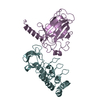
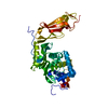
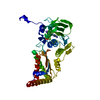
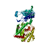
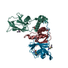
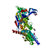
- PDB-1xiw: Crystal structure of human CD3-e/d dimer in complex with a UCHT1 ... -
+ Open data

- Basic information
| Entry | Database: PDB / ID: 1xiw | ||||||
|---|---|---|---|---|---|---|---|
| Title | Crystal structure of human CD3-e/d dimer in complex with a UCHT1 single-chain antibody fragment | ||||||
 Components Components |
| ||||||
 Keywords Keywords | membrane protein/Immune System / CD3-epsilon / CD3-delta / UCHT1-scFv / immunoglobulin fold / antibody-antigen complex / membrane protein-Immune System COMPLEX | ||||||
| Function / homology |  Function and homology information Function and homology informationgamma-delta T cell receptor complex / T cell anergy / positive regulation of cell-cell adhesion mediated by integrin / positive regulation of T cell anergy / CD4-positive, alpha-beta T cell proliferation / gamma-delta T cell activation / negative thymic T cell selection / positive regulation of CD4-positive, alpha-beta T cell proliferation / positive thymic T cell selection / signal complex assembly ...gamma-delta T cell receptor complex / T cell anergy / positive regulation of cell-cell adhesion mediated by integrin / positive regulation of T cell anergy / CD4-positive, alpha-beta T cell proliferation / gamma-delta T cell activation / negative thymic T cell selection / positive regulation of CD4-positive, alpha-beta T cell proliferation / positive thymic T cell selection / signal complex assembly / alpha-beta T cell receptor complex / positive regulation of cell-matrix adhesion / smoothened signaling pathway / T cell receptor complex / Translocation of ZAP-70 to Immunological synapse / Phosphorylation of CD3 and TCR zeta chains / positive regulation of interleukin-4 production / dendrite development / alpha-beta T cell activation / Generation of second messenger molecules / immunological synapse / Co-inhibition by PD-1 / T cell receptor binding / T cell costimulation / positive regulation of calcium-mediated signaling / positive regulation of interleukin-2 production / positive regulation of T cell proliferation / cell surface receptor protein tyrosine kinase signaling pathway / cerebellum development / T cell activation / negative regulation of smoothened signaling pathway / apoptotic signaling pathway / clathrin-coated endocytic vesicle membrane / calcium-mediated signaling / SH3 domain binding / positive regulation of type II interferon production / Immunoregulatory interactions between a Lymphoid and a non-Lymphoid cell / cell-cell junction / transmembrane signaling receptor activity / Downstream TCR signaling / Cargo recognition for clathrin-mediated endocytosis / T cell receptor signaling pathway / Clathrin-mediated endocytosis / signaling receptor complex adaptor activity / cell body / protein-containing complex assembly / regulation of apoptotic process / dendritic spine / adaptive immune response / cell surface receptor signaling pathway / G protein-coupled receptor signaling pathway / negative regulation of gene expression / external side of plasma membrane / positive regulation of gene expression / protein kinase binding / endoplasmic reticulum / Golgi apparatus / identical protein binding / plasma membrane / cytoplasm Similarity search - Function | ||||||
| Biological species |  Homo sapiens (human) Homo sapiens (human) | ||||||
| Method |  X-RAY DIFFRACTION / SYNCHROTRON / MOLECULAR REPLACEMENT / Resolution: 1.9 Å X-RAY DIFFRACTION / SYNCHROTRON / MOLECULAR REPLACEMENT / Resolution: 1.9 Å | ||||||
 Authors Authors | Arnett, K.L. / Harrison, S.C. / Wiley, D.C. | ||||||
 Citation Citation | Journal: Proc.Natl.Acad.Sci.USA / Year: 2004 Title: Crystal structure of a human CD3-epsilon/delta dimer in complex with a UCHT1 single-chain antibody fragment. Authors: Arnett, K.L. / Harrison, S.C. / Wiley, D.C. | ||||||
| History |
| ||||||
| Remark 999 | SEQUENCE The chimera protein consists of immunoglobulin light chain variable region (chains C, G), ...SEQUENCE The chimera protein consists of immunoglobulin light chain variable region (chains C, G), a linker GGGGSGGGGSGGGGS, and immunoglobulin heavy chain variable region (chains D, H). However, the linker GGGGSGGGGSGGGGS are not modeled due to disorder. The conflicts are due to immunoglobulin domain variable region (v) |
- Structure visualization
| Structure viewer | Molecule: MolmilJmol/JSmol |
|---|
- Downloads & links
-Download
| PDBx/mmCIF format | 1xiw.cif.gz | 167 KB | Display | PDBx/mmCIF format |
|---|---|---|---|---|
| PDB format | pdb1xiw.ent.gz | 132.9 KB | Display | PDB format |
| PDBx/mmJSON format | 1xiw.json.gz | Tree view | PDBx/mmJSON format | |
| Others |  Other downloads Other downloads |
-Validation report
| Arichive directory | https://data.pdbj.org/pub/pdb/validation_reports/xi/1xiwftp://data.pdbj.org/pub/pdb/validation_reports/xi/1xiw | HTTPS FTP |
|---|
-Related structure data
| Related structure data | 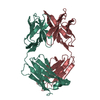 6fabS S: Starting model for refinement |
|---|---|
| Similar structure data |


-Links
 PDBj
PDBj


- Assembly
| Deposited unit | 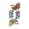
| ||||||||
|---|---|---|---|---|---|---|---|---|---|
| 1 | 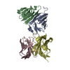
| ||||||||
| 2 | 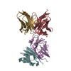
| ||||||||
| Unit cell |
|
-Components
| #1: Protein | Mass: 11893.056 Da / Num. of mol.: 2 / Fragment: ectodomain Source method: isolated from a genetically manipulated source Source: (gene. exp.) Homo sapiens (human) / Gene: CD3E, T3E / Plasmid: pLM1 / Species (production host): Escherichia coli / Production host:  #2: Protein | Mass: 9106.425 Da / Num. of mol.: 2 / Fragment: ectodomain Source method: isolated from a genetically manipulated source Source: (gene. exp.) Homo sapiens (human) / Gene: CD3D, T3D / Plasmid: pLM1 / Species (production host): Escherichia coli / Production host: #3: Antibody | Mass: 11994.338 Da / Num. of mol.: 2 Source method: isolated from a genetically manipulated source Source: (gene. exp.) #4: Antibody | Mass: 13650.221 Da / Num. of mol.: 2 Source method: isolated from a genetically manipulated source Source: (gene. exp.) #5: Water | ChemComp-HOH / |  Mass: 18.015 Da / Num. of mol.: 319 / Source method: isolated from a natural source / Formula: H2O Mass: 18.015 Da / Num. of mol.: 319 / Source method: isolated from a natural source / Formula: H2OHas protein modification | Y | |
|---|
-Experimental details
-Experiment
| Experiment | Method: X-RAY DIFFRACTION / Number of used crystals: 1 |
|---|
- Sample preparation
| Crystal | Density Matthews: 2 Å3/Da / Density % sol: 39.8 % |
|---|---|
| Crystal grow | Temperature: 295 K / Method: vapor diffusion, hanging drop / pH: 7 Details: PEG 3350, sodium chloride, HEPES, pH 7.0, VAPOR DIFFUSION, HANGING DROP, temperature 22K |
-Data collection
| Diffraction | Mean temperature: 100 K |
|---|---|
| Diffraction source | Source: SYNCHROTRON / Site: ALS  / Beamline: 8.2.1 / Wavelength: 0.9796 Å / Beamline: 8.2.1 / Wavelength: 0.9796 Å |
| Detector | Type: ADSC QUANTUM 210 / Detector: CCD / Date: Aug 6, 2003 |
| Radiation | Monochromator: Double crystal, Si(111) / Protocol: SINGLE WAVELENGTH / Monochromatic (M) / Laue (L): M / Scattering type: x-ray |
| Radiation wavelength | Wavelength: 0.9796 Å / Relative weight: 1 |
| Reflection | Resolution: 1.9→50 Å / Num. all: 62308 / Num. obs: 60988 / % possible obs: 97.9 % / Observed criterion σ(F): 0 / Observed criterion σ(I): 0 / Redundancy: 4.9 % / Biso Wilson estimate: 24.8 Å2 / Rmerge(I) obs: 0.065 / Net I/σ(I): 24.4 |
| Reflection shell | Resolution: 1.9→1.97 Å / Redundancy: 5.1 % / Mean I/σ(I) obs: 4.7 / Num. unique all: 6134 / Rsym value: 0.357 / % possible all: 100 |
- Processing
| Software |
| ||||||||||||||||||||||||||||||||||||||||||||||||||||||||||||||||||||||||||||||||
|---|---|---|---|---|---|---|---|---|---|---|---|---|---|---|---|---|---|---|---|---|---|---|---|---|---|---|---|---|---|---|---|---|---|---|---|---|---|---|---|---|---|---|---|---|---|---|---|---|---|---|---|---|---|---|---|---|---|---|---|---|---|---|---|---|---|---|---|---|---|---|---|---|---|---|---|---|---|---|---|---|---|
| Refinement | Method to determine structure: MOLECULAR REPLACEMENT Starting model: PDB ENTRY 6FAB Resolution: 1.9→49.17 Å / Rfactor Rfree error: 0.004 / Data cutoff high absF: 2409958.11 / Data cutoff low absF: 0 / Isotropic thermal model: RESTRAINED / Cross valid method: THROUGHOUT / σ(F): 0 / Stereochemistry target values: Engh & Huber
| ||||||||||||||||||||||||||||||||||||||||||||||||||||||||||||||||||||||||||||||||
| Solvent computation | Solvent model: FLAT MODEL / Bsol: 39.2681 Å2 / ksol: 0.361556 e/Å3 | ||||||||||||||||||||||||||||||||||||||||||||||||||||||||||||||||||||||||||||||||
| Displacement parameters | Biso mean: 30 Å2
| ||||||||||||||||||||||||||||||||||||||||||||||||||||||||||||||||||||||||||||||||
| Refine analyze |
| ||||||||||||||||||||||||||||||||||||||||||||||||||||||||||||||||||||||||||||||||
| Refinement step | Cycle: LAST / Resolution: 1.9→49.17 Å
| ||||||||||||||||||||||||||||||||||||||||||||||||||||||||||||||||||||||||||||||||
| Refine LS restraints |
| ||||||||||||||||||||||||||||||||||||||||||||||||||||||||||||||||||||||||||||||||
| LS refinement shell | Resolution: 1.9→2.02 Å / Rfactor Rfree error: 0.012 / Total num. of bins used: 6
| ||||||||||||||||||||||||||||||||||||||||||||||||||||||||||||||||||||||||||||||||
| Xplor file |
|
