 Movie
Movie Controller

[English] 日本語
 Yorodumi
- PDB-1x06: Crystal structure of undecaprenyl pyrophosphate synthase in compl... -
+ Open data

- Basic information
| Entry | Database: PDB / ID: 1x06 | ||||||
|---|---|---|---|---|---|---|---|
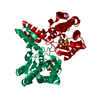
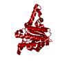
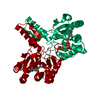
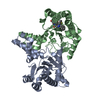
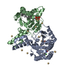
| Title | Crystal structure of undecaprenyl pyrophosphate synthase in complex with Mg, IPP and Fspp | ||||||
 Components Components | Undecaprenyl pyrophosphate synthetase | ||||||
 Keywords Keywords |  TRANSFERASE / enzyme-substrate complex / metal coordination TRANSFERASE / enzyme-substrate complex / metal coordination | ||||||
| Function / homology |  Function and homology information Function and homology informationGram-negative-bacterium-type cell wall biogenesis / ditrans,polycis-undecaprenyl-diphosphate synthase [(2E,6E)-farnesyl-diphosphate specific] / di-trans,poly-cis-undecaprenyl-diphosphate synthase activity / polyprenol biosynthetic process / polyprenyltransferase activity / small molecule binding / peptidoglycan biosynthetic process / cell wall organization / regulation of cell shape / cell cycle ...Gram-negative-bacterium-type cell wall biogenesis / ditrans,polycis-undecaprenyl-diphosphate synthase [(2E,6E)-farnesyl-diphosphate specific] / di-trans,poly-cis-undecaprenyl-diphosphate synthase activity / polyprenol biosynthetic process / polyprenyltransferase activity / small molecule binding / peptidoglycan biosynthetic process / cell wall organization / regulation of cell shape / cell cycle / cell division / magnesium ion binding / protein homodimerization activity / cytosol / cytoplasmSimilarity search - Function | ||||||
| Biological species |  Escherichia coli (E. coli) Escherichia coli (E. coli) | ||||||
| Method | X-RAY DIFFRACTION / SYNCHROTRON / MOLECULAR REPLACEMENT / Resolution: 1.9 Å | ||||||
 Authors Authors | Guo, R.-T. / Ko, T.-P. / Chen, A.P.-C. / Kuo, C.-J. / Wang, A.H.-J. / Liang, P.-H. | ||||||
 Citation Citation | Journal: J.Biol.Chem. / Year: 2005 Title: Crystal structures of undecaprenyl pyrophosphate synthase in complex with magnesium, isopentenyl pyrophosphate, and farnesyl thiopyrophosphate: roles of the metal ion and conserved residues in catalysis. Authors: Guo, R.T. / Ko, T.P. / Chen, A.P. / Kuo, C.J. / Wang, A.H. / Liang, P.H. | ||||||
| History |
|
- Structure visualization
| Structure viewer | Molecule: MolmilJmol/JSmol |
|---|
- Downloads & links
-Download
| PDBx/mmCIF format | 1x06.cif.gz | 69.9 KB | Display | PDBx/mmCIF format |
|---|---|---|---|---|
| PDB format | pdb1x06.ent.gz | 50.1 KB | Display | PDB format |
| PDBx/mmJSON format | 1x06.json.gz | Tree view | PDBx/mmJSON format | |
| Others |  Other downloads Other downloads |
-Validation report
| Arichive directory | https://data.pdbj.org/pub/pdb/validation_reports/x0/1x06ftp://data.pdbj.org/pub/pdb/validation_reports/x0/1x06 | HTTPS FTP |
|---|
-Related structure data
| Related structure data | 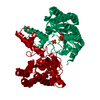 1x07C 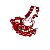 1x08C 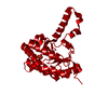 1x09C  1v7uS S: Starting model for refinement C: citing same article ( |
|---|---|
| Similar structure data |


-Links
 PDBj
PDBj- Assembly
| Deposited unit | 
| ||||||||||||
|---|---|---|---|---|---|---|---|---|---|---|---|---|---|
| 1 | 
| ||||||||||||
| Unit cell |
| ||||||||||||
| Components on special symmetry positions |
|
-Components
| #1: Protein | Mass: 28481.127 Da / Num. of mol.: 1 Source method: isolated from a genetically manipulated source Source: (gene. exp.) Escherichia coli (E. coli) / Plasmid: PET32 / Species (production host): Escherichia coli / Production host: Escherichia coli BL21 (bacteria) / Strain (production host): BL21References: UniProt: P60472, ditrans,polycis-undecaprenyl-diphosphate synthase [(2E,6E)-farnesyl-diphosphate specific] | ||
|---|---|---|---|
| #2: Chemical | ChemComp-PO4 / Phosphate  Mass: 94.971 Da / Num. of mol.: 1 / Source method: obtained synthetically / Formula: PO4 Mass: 94.971 Da / Num. of mol.: 1 / Source method: obtained synthetically / Formula: PO4 | ||
| #3: Chemical | ChemComp-MG /   Mass: 24.305 Da / Num. of mol.: 1 / Source method: obtained synthetically / Formula: Mg Mass: 24.305 Da / Num. of mol.: 1 / Source method: obtained synthetically / Formula: Mg | ||
| #4: Chemical |   Mass: 398.392 Da / Num. of mol.: 2 / Source method: obtained synthetically / Formula: C15H28O6P2S Mass: 398.392 Da / Num. of mol.: 2 / Source method: obtained synthetically / Formula: C15H28O6P2S#5: Water | ChemComp-HOH / | Water Mass: 18.015 Da / Num. of mol.: 333 / Source method: isolated from a natural source / Formula: H2O Mass: 18.015 Da / Num. of mol.: 333 / Source method: isolated from a natural source / Formula: H2O |
-Experimental details
-Experiment
| Experiment | Method: X-RAY DIFFRACTION / Number of used crystals: 1 |
|---|
- Sample preparation
| Crystal | Density Matthews: 2.08 Å3/Da / Density % sol: 41 % |
|---|---|
| Crystal grow | Temperature: 298 K / Method: vapor diffusion, hanging drop / pH: 7.5 Details: MgCl2, IPP, FsPP, PEG35000, ethylene glycol, Triton X-100, NaCl, Tris-HCl, pH 7.5, VAPOR DIFFUSION, HANGING DROP, temperature 298K |
-Data collection
| Diffraction | Mean temperature: 100 K |
|---|---|
| Diffraction source | Source: SYNCHROTRON / Site: NSRRC  / Beamline: BL17B2 / Wavelength: 1 Å / Beamline: BL17B2 / Wavelength: 1 Å |
| Detector | Type: RIGAKU RAXIS IV / Detector: IMAGE PLATE / Date: Jul 12, 2004 / Details: mirrors |
| Radiation | Monochromator: Si 111 CHANNEL / Protocol: SINGLE WAVELENGTH / Monochromatic (M) / Laue (L): M / Scattering type: x-ray |
| Radiation wavelength | Wavelength: 1 Å / Relative weight: 1 |
| Reflection | Resolution: 1.9→25 Å / Num. all: 19231 / Num. obs: 19022 / % possible obs: 98.9 % / Observed criterion σ(F): 0 / Observed criterion σ(I): 1 / Redundancy: 4.12 % / Rmerge(I) obs: 0.072 / Net I/σ(I): 19.9 |
| Reflection shell | Resolution: 1.9→1.97 Å / Redundancy: 2.69 % / Rmerge(I) obs: 0.394 / Mean I/σ(I) obs: 2.4 / Num. unique all: 1871 / % possible all: 95 |
- Processing
| Software |
| |||||||||||||||||||||||||
|---|---|---|---|---|---|---|---|---|---|---|---|---|---|---|---|---|---|---|---|---|---|---|---|---|---|---|
| Refinement | Method to determine structure: MOLECULAR REPLACEMENT Starting model: chain A of PDB 1V7U Resolution: 1.9→25 Å / Cross valid method: THROUGHOUT / σ(F): 0 / Stereochemistry target values: Engh & Huber
| |||||||||||||||||||||||||
| Refine analyze |
| |||||||||||||||||||||||||
| Refinement step | Cycle: LAST / Resolution: 1.9→25 Å
| |||||||||||||||||||||||||
| Refine LS restraints |
| |||||||||||||||||||||||||
| LS refinement shell | Resolution: 1.9→1.97 Å / Rfactor Rfree error: 0.018
|
