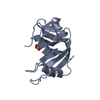
 Movie
Movie Controller


+ Open data

- Basic information
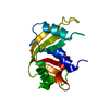
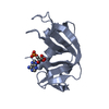
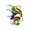
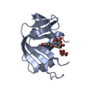
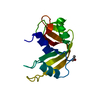
| Entry | Database: PDB / ID: 1rnc | ||||||
|---|---|---|---|---|---|---|---|
| Title | NEWLY OBSERVED BINDING MODE IN PANCREATIC RIBONUCLEASE | ||||||
 Components Components | RIBONUCLEASE A Pancreatic ribonuclease family Pancreatic ribonuclease family | ||||||
 Keywords Keywords | HYDROLASE(ENDORIBONUCLEASE) | ||||||
| Function / homology |  Function and homology informationpancreatic ribonuclease / ribonuclease A activity / RNA nuclease activity / nucleic acid binding / lyase activity / defense response to Gram-positive bacterium / extracellular region Function and homology informationpancreatic ribonuclease / ribonuclease A activity / RNA nuclease activity / nucleic acid binding / lyase activity / defense response to Gram-positive bacterium / extracellular regionSimilarity search - Function | ||||||
| Biological species |  Bos taurus (cattle) Bos taurus (cattle) | ||||||
| Method | X-RAY DIFFRACTION / Resolution: 1.5 Å | ||||||
 Authors Authors | Aguilar, C.F. / Thomas, P.J. / Mills, A. / Moss, D.S. / Palmer, R.A. | ||||||
 Citation Citation | Journal: J.Mol.Biol. / Year: 1992 Title: Newly observed binding mode in pancreatic ribonuclease. Authors: Aguilar, C.F. / Thomas, P.J. / Mills, A. / Moss, D.S. / Palmer, R.A. #1: Journal: Biochim.Biophys.Acta / Year: 1991Title: Novel Non-Productively Bound Ribonuclease Inhibitor Complexes-High Resolution X-Ray Refinement Studies on the Binding of Rnase-A to Cytidylyl-2',5'-Guanosine (2',5'Cpg) and Deoxycytidylyl- ...Title: Novel Non-Productively Bound Ribonuclease Inhibitor Complexes-High Resolution X-Ray Refinement Studies on the Binding of Rnase-A to Cytidylyl-2',5'-Guanosine (2',5'Cpg) and Deoxycytidylyl-3',5'-Guanosine (3',5'Dcpdg) Authors: Aguilar, C.F. / Thomas, P.J. / Moss, D.S. / Mills, A. / Palmer, R.A. #2: Journal: J.Mol.Biol. / Year: 1987Title: X-Ray Refinement Study on the Binding of Cytidylic Acid (2'-Cmp) to Ribonuclease-A Authors: Howlin, B. / Harris, G.W. / Moss, D.S. / Palmer, R.A. #3: Journal: Biochim.Biophys.Acta / Year: 1984Title: An X-Ray Refinement Study on the Binding of Ribonuclease-A to Cytidine-N(3)-Oxide 2'-Phosphate Authors: Palmer, R.A. / Moss, D.S. / Haneef, I. / Borkakoti, N. #4: Journal: J.Crystallogr.Spectrosc.Res. / Year: 1984Title: The Refined Structure of Ribonuclease-A at 1.45 Angstroms Resolution Authors: Borkakoti, N. / Moss, D.S. / Stanford, M.J. / Palmer, R.A. | ||||||
| History |
|
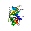
- Structure visualization
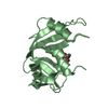
| Structure viewer | Molecule: MolmilJmol/JSmol |
|---|
- Downloads & links
-Download
| PDBx/mmCIF format | 1rnc.cif.gz | 32.8 KB | Display | PDBx/mmCIF format |
|---|---|---|---|---|
| PDB format | pdb1rnc.ent.gz | 25 KB | Display | PDB format |
| PDBx/mmJSON format | 1rnc.json.gz | Tree view | PDBx/mmJSON format | |
| Others |  Other downloads Other downloads |
-Validation report
| Arichive directory | https://data.pdbj.org/pub/pdb/validation_reports/rn/1rncftp://data.pdbj.org/pub/pdb/validation_reports/rn/1rnc | HTTPS FTP |
|---|
-Related structure data

-Links
 PDBj
PDBj


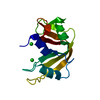
- Assembly
| Deposited unit | 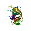
| ||||||||
|---|---|---|---|---|---|---|---|---|---|
| 1 |
| ||||||||
| Unit cell |
| ||||||||
| Atom site foot note | 1: RESIDUES PRO 93 AND PRO 114 ARE CIS PROLINES. 2: PEPTIDE BOND DEVIATES SIGNIFICANTLY FROM TRANS CONFORMATION GLU 111 - GLY 112: 149.724 |
-Components
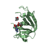
| #1: Protein | Pancreatic ribonuclease family Mass: 13708.326 Da / Num. of mol.: 1 Source method: isolated from a genetically manipulated source Source: (gene. exp.) Bos taurus (cattle) / Organ: PANCREAS / References: UniProt: P61823, EC: 3.1.27.5 |
|---|---|
| #2: Chemical | ChemComp-SO4 / Sulfate  Mass: 96.063 Da / Num. of mol.: 1 / Source method: obtained synthetically / Formula: SO4 Mass: 96.063 Da / Num. of mol.: 1 / Source method: obtained synthetically / Formula: SO4 |
| #3: Chemical | ChemComp-5GP / Guanosine monophosphate  Mass: 363.221 Da / Num. of mol.: 1 / Source method: obtained synthetically / Formula: C10H14N5O8P Mass: 363.221 Da / Num. of mol.: 1 / Source method: obtained synthetically / Formula: C10H14N5O8P |
| #4: Water | ChemComp-HOH / Water Mass: 18.015 Da / Num. of mol.: 30 / Source method: isolated from a natural source / Formula: H2O Mass: 18.015 Da / Num. of mol.: 30 / Source method: isolated from a natural source / Formula: H2O |
| Compound details | THE MOST INTERESTING FEATURE IN THIS STRUCTURE IS THE MODE OF BINDING OF THE CPG INHIBITOR TO THE ...THE MOST INTERESTIN |
-Experimental details
-Experiment
| Experiment | Method: X-RAY DIFFRACTION |
|---|
- Sample preparation
| Crystal | Density Matthews: 2.18 Å3/Da / Density % sol: 43.61 % |
|---|---|
| Crystal grow | *PLUS Method: unknown / PH range low: 5.7 / PH range high: 5.2 |
| Components of the solutions | *PLUS Conc.: 47 %(v/v) / Common name: ethanol |
-Data collection
| Radiation | Scattering type: x-ray |
|---|---|
| Radiation wavelength | Relative weight: 1 |
- Processing
| Software |
| ||||||||||||
|---|---|---|---|---|---|---|---|---|---|---|---|---|---|
| Refinement | Highest resolution: 1.5 Å Details: THE INHIBITOR AND SULFATE ANION WERE REFINED USING GROUP OCCUPANCIES.
| ||||||||||||
| Refinement step | Cycle: LAST / Highest resolution: 1.5 Å
| ||||||||||||
| Refine LS restraints |
| ||||||||||||
| Refinement | *PLUS Highest resolution: 1.5 Å / Num. reflection obs: 17855 / Rfactor obs: 0.21 | ||||||||||||
| Solvent computation | *PLUS | ||||||||||||
| Displacement parameters | *PLUS | ||||||||||||
| Refine LS restraints | *PLUS Type: p_angle_d / Dev ideal: 1.23 |
