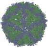
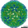
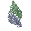
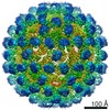
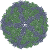
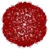
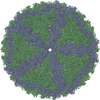
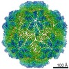
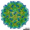
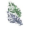
ジャーナル: J Virol / 年: 2021 タイトル: Capsid Structure of RNA Virus 1. 著者: Michaela Procházková / Tibor Füzik / Danyil Grybchuk / Francesco Luca Falginella / Lucie Podešvová / Vyacheslav Yurchenko / Robert Vácha / Pavel Plevka / 要旨: parasites cause a variety of symptoms, including mucocutaneous leishmaniasis, which results in the destruction of the mucous membranes of the nose, mouth, and throat. The species of carrying RNA ... parasites cause a variety of symptoms, including mucocutaneous leishmaniasis, which results in the destruction of the mucous membranes of the nose, mouth, and throat. The species of carrying RNA virus 1 (LRV1), from the family , are more likely to cause severe disease and are less sensitive to treatment than those that do not contain the virus. Although the importance of LRV1 for the severity of leishmaniasis was discovered a long time ago, the structure of the virus remained unknown. Here, we present a cryo-electron microscopy reconstruction of the virus-like particle of LRV1 determined to a resolution of 3.65 Å. The capsid has icosahedral symmetry and is formed by 120 copies of a capsid protein assembled in asymmetric dimers. RNA genomes of viruses from the family are synthetized, but not capped at the 5' end, by virus RNA polymerases. To protect viral RNAs from degradation, capsid proteins of the L-A totivirus cleave the 5' caps of host mRNAs, creating decoys to overload the cellular RNA quality control system. Capsid proteins of LRV1 form positively charged clefts, which may be the cleavage sites for the 5' cap of mRNAs. The putative RNA binding site of LRV1 is distinct from that of the related L-A virus. The structure of the LRV1 capsid enables the rational design of compounds targeting the putative decapping site. Such inhibitors may be developed into a treatment for mucocutaneous leishmaniasis caused by LRV1-positive species of Twelve million people worldwide suffer from leishmaniasis, resulting in more than 30 thousand deaths annually. The disease has several variants that differ in their symptoms. The mucocutaneous form, which leads to disintegration of the nasal septum, lips, and palate, is caused predominantly by parasites carrying RNA virus 1 (LRV1). Here, we present the structure of the LRV1 capsid determined using cryo-electron microscopy. Capsid proteins of a related totivirus, L-A virus, protect viral RNAs from degradation by cleaving the 5' caps of host mRNAs. Capsid proteins of LRV1 may have the same function. We show that the LRV1 capsid contains positively charged clefts that may be sites for the cleavage of mRNAs of cells. The structure of the LRV1 capsid enables the rational design of compounds targeting the putative mRNA cleavage site. Such inhibitors may be used as treatments for mucocutaneous leishmaniasis.
分子量: 87536.930 Da / 分子数: 2 / 由来タイプ: 組換発現 詳細: Subunit A model for residues 15-204, 210-520, 541-635. Subunit B model for residues 19-290, 300-517, 541-576, 583-642.,Subunit A model for residues 15-204, 210-520, 541-635. Subunit B model ...詳細: Subunit A model for residues 15-204, 210-520, 541-635. Subunit B model for residues 19-290, 300-517, 541-576, 583-642.,Subunit A model for residues 15-204, 210-520, 541-635. Subunit B model for residues 19-290, 300-517, 541-576, 583-642. 由来: (組換発現) Leishmania RNA virus 1 - 4 (ウイルス) プラスミド: pET42b / 詳細 (発現宿主): C-terminal 8xHis tag / 発現宿主: Escherichia coli BL21(DE3) (大腸菌) / 参照: UniProt: L7XUU7
 ムービー
ムービー コントローラー


 データを開く
 基本情報
基本情報 要素
要素 カプシド
カプシド  キーワード
キーワード 機能・相同性情報
 Leishmania RNA virus 1 - 4 (ウイルス)
 データ登録者
 チェコ, 1件
  引用
引用
 構造の表示
構造の表示 ダウンロードとリンク
 その他のダウンロード
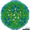

 PDBj
PDBj 集合体
集合体

 試料調製
試料調製 電子顕微鏡撮影
 解析
解析