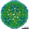
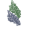
Journal: Virology / Year: 2022 Title: Virion structure of Leishmania RNA virus 1. Authors: Michaela Procházková / Tibor Füzik / Danyil Grybchuk / Vyacheslav Yurchenko / Pavel Plevka / Abstract: The presence of Leishmania RNA virus 1 (LRV1) enables Leishmania protozoan parasites to cause more severe disease than the virus-free strains. The structure of LRV1 virus-like particles has been ...The presence of Leishmania RNA virus 1 (LRV1) enables Leishmania protozoan parasites to cause more severe disease than the virus-free strains. The structure of LRV1 virus-like particles has been determined previously, however, the structure of the LRV1 virion has not been characterized. Here we used cryo-electron microscopy and single-particle reconstruction to determine the structures of the LRV1 virion and empty particle isolated from Leishmania guyanensis to resolutions of 4.0 Å and 3.6 Å, respectively. The capsid of LRV1 is built from sixty dimers of capsid proteins organized with icosahedral symmetry. RNA genomes of totiviruses are replicated inside the virions by RNA polymerases expressed as C-terminal extensions of a sub-population of capsid proteins. Most of the virions probably contain one or two copies of the RNA polymerase, however, the location of the polymerase domains in LRV1 capsid could not be identified, indicating that it varies among particles. Importance. Every year over 200 000 people contract leishmaniasis and more than five hundred people die of the disease. The mucocutaneous form of leishmaniasis produces lesions that can destroy the mucous membranes of the nose, mouth, and throat. Leishmania parasites carrying Leishmania RNA virus 1 (LRV1) are predisposed to cause aggravated symptoms in the mucocutaneous form of leishmaniasis. Here, we present the structure of the LRV1 virion determined using cryo-electron microscopy.
Electron dose: 1.3 e/Å2 / Detector mode: SUPER-RESOLUTION / Film or detector model: GATAN K2 SUMMIT (4k x 4k) / Num. of grids imaged: 1 / Num. of real images: 3981
Image scans
Movie frames/image: 40
-
Processing
EM software
ID
Name
Version
Category
1
crYOLO
particleselection
2
EPU
imageacquisition
4
CTFFIND
4
CTFcorrection
7
Coot
0.8.9.2
modelfitting
9
PHENIX
1.15.2
modelrefinement
12
RELION
classification
13
RELION
3.7
3Dreconstruction
CTF correction
Type: PHASE FLIPPING AND AMPLITUDE CORRECTION
Particle selection
Num. of particles selected: 7912
Symmetry
Point symmetry: I (icosahedral)
3D reconstruction
Resolution: 3.63 Å / Resolution method: FSC 0.143 CUT-OFF / Num. of particles: 3768 / Algorithm: FOURIER SPACE / Num. of class averages: 1 / Symmetry type: POINT
Atomic model building
B value: 72.61 / Protocol: RIGID BODY FIT / Space: RECIPROCAL / Target criteria: correlation coefficient
In the structure databanks used in Yorodumi, some data are registered as the other names, "COVID-19 virus" and "2019-nCoV". Here are the details of the virus and the list of structure data.
Jan 31, 2019. EMDB accession codes are about to change! (news from PDBe EMDB page)
EMDB accession codes are about to change! (news from PDBe EMDB page)
The allocation of 4 digits for EMDB accession codes will soon come to an end. Whilst these codes will remain in use, new EMDB accession codes will include an additional digit and will expand incrementally as the available range of codes is exhausted. The current 4-digit format prefixed with “EMD-” (i.e. EMD-XXXX) will advance to a 5-digit format (i.e. EMD-XXXXX), and so on. It is currently estimated that the 4-digit codes will be depleted around Spring 2019, at which point the 5-digit format will come into force.
The EM Navigator/Yorodumi systems omit the EMD- prefix.
Related info.:Q: What is EMD? / ID/Accession-code notation in Yorodumi/EM Navigator
Yorodumi is a browser for structure data from EMDB, PDB, SASBDB, etc.
This page is also the successor to EM Navigator detail page, and also detail information page/front-end page for Omokage search.
The word "yorodu" (or yorozu) is an old Japanese word meaning "ten thousand". "mi" (miru) is to see.
Related info.:EMDB / PDB / SASBDB / Comparison of 3 databanks / Yorodumi Search / Aug 31, 2016. New EM Navigator & Yorodumi / Yorodumi Papers / Jmol/JSmol / Function and homology information / Changes in new EM Navigator and Yorodumi
 Movie
Movie Controller


 Open data
 Basic information
 Components
 Keywords
 Function and homology information
 Leishmania RNA virus 1 - LgM5313
 Authors
 Czech Republic, 1items
  Citation
 Structure visualization
 Downloads & links
 Other downloads
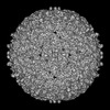


 PDBj
PDBj Assembly
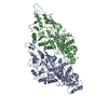
 Sample preparation
 Electron microscopy imaging
 FIELD EMISSION GUN / Accelerating voltage: 300 kV / Illumination mode: FLOOD BEAM
 Processing