| 登録情報 | データベース: PDB / ID: 4duu
|
|---|
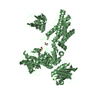
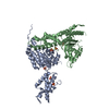
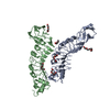
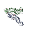
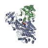
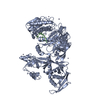
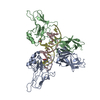
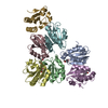
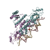
| タイトル | The X-ray Crystal Structure of Full-Length type I Human Plasminogen |
|---|
 要素 要素 | Plasminogen プラスミン プラスミン |
|---|
 キーワード キーワード | HYDROLASE (加水分解酵素) / serine protease (セリンプロテアーゼ) / fibrinolysis |
|---|
| 機能・相同性 |  機能・相同性情報 機能・相同性情報
プラスミン / trans-synaptic signaling by BDNF, modulating synaptic transmission / trophoblast giant cell differentiation / tissue remodeling / protein antigen binding / 再生 (生物学) / mononuclear cell migration / Signaling by PDGF / negative regulation of cell-cell adhesion mediated by cadherin / positive regulation of fibrinolysis ...プラスミン / trans-synaptic signaling by BDNF, modulating synaptic transmission / trophoblast giant cell differentiation / tissue remodeling / protein antigen binding / 再生 (生物学) / mononuclear cell migration / Signaling by PDGF / negative regulation of cell-cell adhesion mediated by cadherin / positive regulation of fibrinolysis / Dissolution of Fibrin Clot / negative regulation of cell-substrate adhesion / myoblast differentiation / biological process involved in interaction with symbiont / labyrinthine layer blood vessel development / muscle cell cellular homeostasis / Activation of Matrix Metalloproteinases / apolipoprotein binding / extracellular matrix disassembly / positive regulation of blood vessel endothelial cell migration / negative regulation of fibrinolysis / fibrinolysis / Degradation of the extracellular matrix / serine-type peptidase activity / platelet alpha granule lumen / Schaffer collateral - CA1 synapse / kinase binding / Regulation of Insulin-like Growth Factor (IGF) transport and uptake by Insulin-like Growth Factor Binding Proteins (IGFBPs) / 凝固・線溶系 / Platelet degranulation / protein-folding chaperone binding / collagen-containing extracellular matrix / blood microparticle / endopeptidase activity / protease binding / protein domain specific binding / negative regulation of cell population proliferation / external side of plasma membrane / signaling receptor binding / serine-type endopeptidase activity / glutamatergic synapse / enzyme binding / 細胞膜 / タンパク質分解 / extracellular space / extracellular exosome / extracellular region / 細胞膜類似検索 - 分子機能 Peptidase S1A, plasmin / divergent subfamily of APPLE domains / PAN/Apple domain profile. / PAN domain / PAN/Apple domain / Kringle domain / Kringle / Kringle, conserved site / Kringle superfamily / Kringle domain signature. ...Peptidase S1A, plasmin / divergent subfamily of APPLE domains / PAN/Apple domain profile. / PAN domain / PAN/Apple domain / Kringle domain / Kringle / Kringle, conserved site / Kringle superfamily / Kringle domain signature. / Kringle domain profile. / Kringle domain / Kringle-like fold / Serine proteases, trypsin family, histidine active site / Serine proteases, trypsin family, serine active site / Peptidase S1A, chymotrypsin family / Serine proteases, trypsin family, histidine active site. / Serine proteases, trypsin domain profile. / Serine proteases, trypsin family, serine active site. / Trypsin-like serine protease / Serine proteases, trypsin domain / トリプシン / Peptidase S1, PA clan, chymotrypsin-like fold / Peptidase S1, PA clan類似検索 - ドメイン・相同性 |
|---|
| 生物種 |  Homo sapiens (ヒト) Homo sapiens (ヒト) |
|---|
| 手法 | X線回折 / シンクロトロン / 分子置換 / 解像度: 5.2 Å |
|---|
 データ登録者 データ登録者 | Law, R.H.P. / Caradoc-Davies, T. / Whisstock, J.C. |
|---|
 引用 引用 | ジャーナル: Cell Rep / 年: 2012
タイトル: The X-ray crystal structure of full-length human plasminogen
著者: Law, R.H.P. / Caradoc-Davies, T. / Cowieson, N. / Horvath, A.J. / Quek, A.J. / Encarnacao, J.A. / Steer, D. / Cowan, A. / Zhang, Q. / Lu, B.G.C. / Pike, R.N. / Smith, A.I. / Coughlin, P.B. / Whisstock, J.C. |
|---|
| 履歴 | | 登録 | 2012年2月22日 | 登録サイト: RCSB / 処理サイト: PDBJ |
|---|
| 改定 1.0 | 2012年3月28日 | Provider: repository / タイプ: Initial release |
|---|
| 改定 1.1 | 2012年4月4日 | Group: Structure summary |
|---|
| 改定 1.2 | 2013年6月26日 | Group: Database references |
|---|
| 改定 1.3 | 2023年11月8日 | Group: Data collection / Database references / Refinement description
カテゴリ: chem_comp_atom / chem_comp_bond ...chem_comp_atom / chem_comp_bond / database_2 / pdbx_initial_refinement_model
Item: _database_2.pdbx_DOI / _database_2.pdbx_database_accession |
|---|
|
|---|
 ムービー
ムービー コントローラー


 データを開く
 基本情報
基本情報 構造の表示
構造の表示 ダウンロードとリンク
 その他のダウンロード
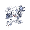
 PDBj
PDBj


 集合体
集合体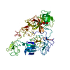
 分子量: 18.015 Da / 分子数: 35 / 由来タイプ: 天然 / 式: H2O
 試料調製
試料調製 / ビームライン: MX2 / 波長: 0.953697 Å
 解析
解析