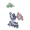
 ムービー
ムービー コントローラー


+ データを開く

- 基本情報
基本情報
| 登録情報 | データベース: PDB / ID: 1jqm | ||||||
|---|---|---|---|---|---|---|---|
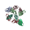
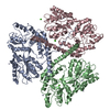
| タイトル | Fitting of L11 protein and elongation factor G (EF-G) in the cryo-em map of e. coli 70S ribosome bound with EF-G, GDP and fusidic acid | ||||||
 要素 要素 |
| ||||||
 キーワード キーワード |  RIBOSOME (リボソーム) / L11 / EF-G (EF-G) / cryo-EM (低温電子顕微鏡法) / 70s E.coli ribosome / GDP state RIBOSOME (リボソーム) / L11 / EF-G (EF-G) / cryo-EM (低温電子顕微鏡法) / 70s E.coli ribosome / GDP state | ||||||
| 機能・相同性 |  機能・相同性情報 機能・相同性情報translational elongation / translation elongation factor activity / GDP binding / large ribosomal subunit rRNA binding / ribosome binding / cytosolic large ribosomal subunit / structural constituent of ribosome / 翻訳 (生物学) / GTPase activity / GTP binding ...translational elongation / translation elongation factor activity / GDP binding / large ribosomal subunit rRNA binding / ribosome binding / cytosolic large ribosomal subunit / structural constituent of ribosome / 翻訳 (生物学) / GTPase activity / GTP binding / magnesium ion binding / 細胞質類似検索 - 分子機能 | ||||||
| 生物種 |  Escherichia coli (大腸菌) Escherichia coli (大腸菌) | ||||||
| 手法 | 電子顕微鏡法 / 単粒子再構成法 / Molecular Modeling based on crystal structures / クライオ電子顕微鏡法 / 解像度: 18 Å | ||||||
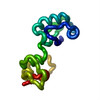
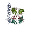
| Model details | This structure was generated by fitting the X-ray crystal structure of L11 and EF-G into the 70S E. ...This structure was generated by fitting the X-ray crystal structure of L11 and EF-G into the 70S E. coli EF-G- (GDP form) bound ribosome map. L11 (linker region between N and C terminal) and EF-G positions of domains G' and V were modeled to accommodate the conformational changes. The structure is modeled as a C-alpha trace. Conformational changes occur in protein L11 and EF-G in two stpes due to the binding of EF-G to the 70S ribosome, and subsequent GTP hydrolysis. These changed conformations were modeled based on the fitting of the crystal coordinates to the low resolution ribosome map (factor-bound) and energy minimizing the fitted structures. | ||||||
 データ登録者 データ登録者 | Agrawal, R.K. / Linde, J. / Segupta, J. / Nierhaus, K.H. / Frank, J. | ||||||
 引用 引用 | ジャーナル: J Mol Biol / 年: 2001 タイトル: Localization of L11 protein on the ribosome and elucidation of its involvement in EF-G-dependent translocation. 著者: R K Agrawal / J Linde / J Sengupta / K H Nierhaus / J Frank /  要旨: L11 protein is located at the base of the L7/L12 stalk of the 50 S subunit of the Escherichia coli ribosome. Because of the flexible nature of the region, recent X-ray crystallographic studies of the ...L11 protein is located at the base of the L7/L12 stalk of the 50 S subunit of the Escherichia coli ribosome. Because of the flexible nature of the region, recent X-ray crystallographic studies of the 50 S subunit failed to locate the N-terminal domain of the protein. We have determined the position of the complete L11 protein by comparing a three-dimensional cryo-EM reconstruction of the 70 S ribosome, isolated from a mutant lacking ribosomal protein L11, with the three-dimensional map of the wild-type ribosome. Fitting of the X-ray coordinates of L11-23 S RNA complex and EF-G into the cryo-EM maps combined with molecular modeling, reveals that, following EF-G-dependent GTP hydrolysis, domain V of EF-G intrudes into the cleft between the 23 S ribosomal RNA and the N-terminal domain of L11 (where the antibiotic thiostrepton binds), causing the N-terminal domain to move and thereby inducing the formation of the arc-like connection with the G' domain of EF-G. The results provide a new insight into the mechanism of EF-G-dependent translocation. #1: ジャーナル: Cell(Cambridge,Mass.) / 年: 1999タイトル: A Detailed View of a Ribosomal Active Site: The Structure of the L11-RNA Complex 著者: Wimberly, B.T. / Guymon, R. / McCutcheon, J.P. / White, S.W. / Ramakrishnan, V. #2: ジャーナル: Embo J. / 年: 1994タイトル: The Crystal Structure of Elongation Factor G Complexed with GDP, at 2.7A Resolution. 著者: Czworkowski, J. / Wang, J. / Steitz, T.A. / Moore, P.B. #3: ジャーナル: Embo J. / 年: 1994タイトル: Three-dimensional structure of the ribosomal translocase: Elongation factor G from Thermus thermophilus 著者: AEvarsson, A. / Brazhnikov, E. / Garber, M. / Zheltonosova, J. / Chirgadze, Y. / al-Karadaghi, S. / Svensson, L.A. / Liljas, A. #4: ジャーナル: J.Mol.Biol. / 年: 2000タイトル: Structure of a Mutant EF-G Reveals Domain III and Possibly the Fusidic Acid Binding Site 著者: Laurberg, M. / Kristensen, O. / Martemyanov, K. / Gudkov, A.T. / Nagaev, I. / Hughes, D. / Liljas, A. #5: ジャーナル: Nat.Struct.Biol. / 年: 1999タイトル: EF-G-dependent GTP hydrolysis induces translocation accompanied by large conformational changes in the 70S ribosome 著者: Agrawal, R.K. / Heagle, A.B. / Penczek, P. / Grassucci, R.A. / Frank, J. | ||||||
| 履歴 |
|
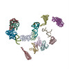
- 構造の表示
構造の表示
| ムービー |
ムービービューア |
|---|---|
| 構造ビューア | 分子: MolmilJmol/JSmol |
- ダウンロードとリンク
-ダウンロード
| PDBx/mmCIF形式 | 1jqm.cif.gz | 39.5 KB | 表示 | PDBx/mmCIF形式 |
|---|---|---|---|---|
| PDB形式 | pdb1jqm.ent.gz | 20.3 KB | 表示 | PDB形式 |
| PDBx/mmJSON形式 | 1jqm.json.gz | ツリー表示 | PDBx/mmJSON形式 | |
| その他 |  その他のダウンロード その他のダウンロード |
-検証レポート
| アーカイブディレクトリ | https://data.pdbj.org/pub/pdb/validation_reports/jq/1jqmftp://data.pdbj.org/pub/pdb/validation_reports/jq/1jqm | HTTPS FTP |
|---|
-関連構造データ
-リンク
 PDBj
PDBj


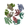
- 集合体
集合体
| 登録構造単位 | 
|
|---|---|
| 1 |
|
-要素
| #1: タンパク質 | / 座標モデル: Cα原子のみ 分子量: 14865.637 Da / 分子数: 1 / 由来タイプ: 天然 詳細: L11 from E. coli 70S ribosome modeled by crystal structure of L11 from Thermatogoma maritima 由来: (天然) Escherichia coli (大腸菌) / 参照: UniProt: P29395 |
|---|---|
| #2: タンパク質 | EF-G / EF-G / translation elongation factor EF-G / 座標モデル: Cα原子のみ 分子量: 76963.078 Da / 分子数: 1 / 由来タイプ: 天然 詳細: EF-G from E. coli 70S ribosome modeled by crystal structure of EF-G from Thermus thermophilus 由来: (天然) Escherichia coli (大腸菌) / 参照: UniProt: P13551 |
-実験情報
-実験
| 実験 | 手法: 電子顕微鏡法 |
|---|---|
| EM実験 | 試料の集合状態: PARTICLE / 3次元再構成法: 単粒子再構成法 |
| NMR実験の詳細 | Text: This structure was generated by fitting the X-ray crystal structure of L11 and EF-G into the 70S E. coli EF-G (GDP form) bound ribosome map. L11 (linker region between N and C terminal) and EF- ...Text: This structure was generated by fitting the X-ray crystal structure of L11 and EF-G into the 70S E. coli EF-G (GDP form) bound ribosome map. L11 (linker region between N and C terminal) and EF-G positions of domains G' and V were modeled to accommodate the conformational changes. |
- 試料調製
試料調製
| 構成要素 | 名称: e. coli 70S ribosome bound with EF-G, GDP and fusidic acid タイプ: RIBOSOME |
|---|---|
| 緩衝液 | pH: 7.6 |
| 試料 | 包埋: NO / シャドウイング: NO / 染色: NO / 凍結: YES |
| 結晶化 | *PLUS 手法: 電子顕微鏡法 |
- 電子顕微鏡撮影
| 顕微鏡 | モデル: FEI/PHILIPS EM420 |
|---|---|
| 電子銃 | 電子線源: その他 / 照射モード: FLOOD BEAM |
| 電子レンズ | モード: BRIGHT FIELDBright-field microscopy / 倍率(公称値): 52000 X |
| 試料ホルダ | 試料ホルダーモデル: GATAN LIQUID NITROGEN |
| 撮影 | 電子線照射量: 10 e/Å2 / フィルム・検出器のモデル: GENERIC FILM |
- 解析
解析
| 粒子像の選択 | 選択した粒子像数: 36113 | ||||||||||||||||||||
|---|---|---|---|---|---|---|---|---|---|---|---|---|---|---|---|---|---|---|---|---|---|
| 対称性 | 点対称性: C1 (非対称) | ||||||||||||||||||||
| 3次元再構成 | 解像度: 18 Å / 解像度の算出法: FSC 0.5 CUT-OFF / 粒子像の数: 36113 / 対称性のタイプ: POINT | ||||||||||||||||||||
| 原子モデル構築 | プロトコル: OTHER / 空間: REAL / Target criteria: VISUAL AGREEMENT 詳細: REFINEMENT PROTOCOL--MANUAL DETAILS--This structure was generated by fitting the X-ray crystal structure of L11 and EF-G into the 70S E. coli EF-G (GDP form) bound ribosome electron ...詳細: REFINEMENT PROTOCOL--MANUAL DETAILS--This structure was generated by fitting the X-ray crystal structure of L11 and EF-G into the 70S E. coli EF-G (GDP form) bound ribosome electron microscopy map. L11 (linker region between N and C terminal) and EF-G positions of domains G and V were modeled to accommodate the conformational changes. Conformational changes occur in protein L11 and EF-G in two steps due to the binding of EF-G to the 70S ribosome, and subsequent GTP hydrolysis. These changed conformations were modeled based on the fitting of the crystal coordinates to the low resolution ribosome map (factor-bound) and energy minimizing the fitted structures. | ||||||||||||||||||||
| 精密化ステップ | サイクル: LAST
| ||||||||||||||||||||
| NMR software |
| ||||||||||||||||||||
| 精密化 | 手法: Molecular Modeling based on crystal structures / ソフトェア番号: 1 詳細: Conformational changes occur in protein L11 and EF-G in two stpes due to the binding of EF-G to the 70S ribosome, and subsequent GTP hydrolysis. These changed conformations were modeled based ...詳細: Conformational changes occur in protein L11 and EF-G in two stpes due to the binding of EF-G to the 70S ribosome, and subsequent GTP hydrolysis. These changed conformations were modeled based on the fitting of the crystal coordinates to the low resolution ribosome map (factor-bound) and energy minimizing the fitted structures. |
