- EMDB-8818: Cryo-EM structure of the P73G mutant of cucumber necrosis virus u... -
+
データを開く
IDまたはキーワード:
読み込み中...
-
基本情報
登録情報
データベース: EMDB / ID: EMD-8818
タイトル
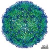
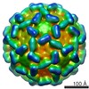
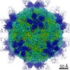
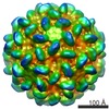
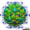
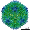
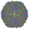
Cryo-EM structure of the P73G mutant of cucumber necrosis virus under native conditions
マップデータ
Cucumber necrosis virus, P73G mutant. Mutation blocks insect vector transmission but is buried in the capsid. Map shows disruption in the putative Zn binding site that may explain loss of transmission.
試料
ウイルス: Cucumber necrosis virus (ウイルス)
機能・相同性
Plant viruses icosahedral capsid proteins 'S' region signature. / Icosahedral viral capsid protein, S domain / Viral coat protein (S domain) / T=3 icosahedral viral capsid / Viral coat protein subunit / structural molecule activity / RNA binding / Capsid protein
ジャーナル: J Virol / 年: 2017 タイトル: Stability of Cucumber Necrosis Virus at the Quasi-6-Fold Axis Affects Zoospore Transmission. 著者: Michael B Sherman / Kishore Kakani / D'Ann Rochon / Wen Jiang / Neil R Voss / Thomas J Smith / 要旨: (CNV) is a member of the genus and has a monopartite positive-sense RNA genome. CNV is transmitted in nature via zoospores of the fungus As with other members of the genus, the CNV capsid swells ... (CNV) is a member of the genus and has a monopartite positive-sense RNA genome. CNV is transmitted in nature via zoospores of the fungus As with other members of the genus, the CNV capsid swells when exposed to alkaline pH and EDTA. We previously demonstrated that a P73G mutation blocks the virus from zoospore transmission while not significantly affecting replication in plants (K. Kakani, R. Reade, and D. Rochon, J Mol Biol 338:507-517, 2004, https://doi.org/10.1016/j.jmb.2004.03.008). P73 lies immediately adjacent to a putative zinc binding site (M. Li et al., J Virol 87:12166-12175, 2013, https://doi.org/10.1128/JVI.01965-13) that is formed by three icosahedrally related His residues in the N termini of the C subunit at the quasi-6-fold axes. To better understand how this buried residue might affect vector transmission, we determined the cryo-electron microscopy structure of wild-type CNV in the native and swollen state and of the transmission-defective mutant, P73G, under native conditions. With the wild-type CNV, the swollen structure demonstrated the expected expansion of the capsid. However, the zinc binding region at the quasi-6-fold at the β-annulus axes remained intact. By comparison, the zinc binding region of the P73G mutant, even under native conditions, was markedly disordered, suggesting that the β-annulus had been disrupted and that this could destabilize the capsid. This was confirmed with pH and urea denaturation experiments in conjunction with electron microscopy analysis. We suggest that the P73G mutation affects the zinc binding and/or the β-annulus, making it more fragile under neutral/basic pH conditions. This, in turn, may affect zoospore transmission. (CNV), a member of the genus , is transmitted in nature via zoospores of the fungus While a number of plant viruses are transmitted via insect vectors, little is known at the molecular level as to how the viruses are recognized and transmitted. As with many spherical plant viruses, the CNV capsid swells when exposed to alkaline pH and EDTA. We previously demonstrated that a P73G mutation that lies inside the capsid immediately adjacent to a putative zinc binding site (Li et al., J Virol 87:12166-12175, 2013, https://doi.org/10.1128/JVI.01965-13) blocks the virus from zoospore transmission while not significantly affecting replication in plants (K. Kakani, R. Reade, and D. Rochon, J Mol Biol 338:507-517, 2004, https://doi.org/10.1016/j.jmb.2004.03.008). Here, we show that the P73G mutant is less stable than the wild type, and this appears to be correlated with destabilization of the β-annulus at the icosahedral 3-fold axes. Therefore, the β-annulus appears not to be essential for particle assembly but is necessary for interactions with the transmission vector.
#262 - 2021年10月 PDB構造への50年間のオープンアクセス (Fifty Years of Open Access to PDB Structures ) 類似性 (3)
#200 - 2016年8月 正二十面体型ウイルスの準対称性 (Quasisymmetry in Icosahedral Viruses) 類似性 (4)
-
マップ
ファイル
ダウンロード / ファイル: emd_8818.map.gz / 形式: CCP4 / 大きさ: 634.7 MB / タイプ: IMAGE STORED AS FLOATING POINT NUMBER (4 BYTES)
注釈
Cucumber necrosis virus, P73G mutant. Mutation blocks insect vector transmission but is buried in the capsid. Map shows disruption in the putative Zn binding site that may explain loss of transmission.
 ムービー
ムービー コントローラー


 データを開く
 基本情報
基本情報 マップデータ
 試料
試料 機能・相同性情報
 Cucumber necrosis virus (ウイルス)
 データ登録者
 引用
引用

 構造の表示
構造の表示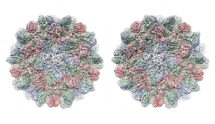
 ダウンロードとリンク
 emd_8818.png
 http://ftp.pdbj.org/pub/emdb/structures/EMD-8818


 試料の構成要素
 解析
解析 電子顕微鏡法
 FIELD EMISSION GUN