 ムービー
ムービー コントローラー


+ データを開く

- 基本情報
基本情報
| 登録情報 | データベース: EMDB / ID: EMD-8806 | |||||||||
|---|---|---|---|---|---|---|---|---|---|---|
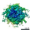
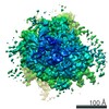
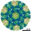
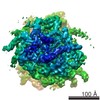
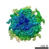
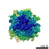
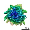
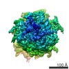
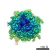
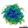
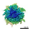
| タイトル | 80S ribsome from Oryctolagus cuniculus, class V - with mid-rotated/swiveled 40S and eeF2, derived from EMPIAR-10064 using eClarity | |||||||||
 マップデータ マップデータ | Rabbit 80S ribosome derived from EMPIAR-10064 using emClarity. Mid-rotated/swiveled 40S with eeF2 | |||||||||
 試料 試料 |
| |||||||||
| 生物種 |   Oryctolagus cuniculus (ウサギ) Oryctolagus cuniculus (ウサギ) | |||||||||
| 手法 | サブトモグラム平均法 / クライオ電子顕微鏡法 / 解像度: 11.0 Å | |||||||||
 データ登録者 データ登録者 | Himes BA / Zhang P | |||||||||
| 資金援助 |  米国, 1件 米国, 1件
| |||||||||
 引用 引用 | ジャーナル: Nat Methods / 年: 2018 タイトル: emClarity: software for high-resolution cryo-electron tomography and subtomogram averaging. 著者: Benjamin A Himes / Peijun Zhang /  要旨: Macromolecular complexes are intrinsically flexible and often challenging to purify for structure determination by single-particle cryo-electron microscopy (cryo-EM). Such complexes can be studied by ...Macromolecular complexes are intrinsically flexible and often challenging to purify for structure determination by single-particle cryo-electron microscopy (cryo-EM). Such complexes can be studied by cryo-electron tomography (cryo-ET) combined with subtomogram alignment and classification, which in exceptional cases achieves subnanometer resolution, yielding insight into structure-function relationships. However, it remains challenging to apply this approach to specimens that exhibit conformational or compositional heterogeneity or are present in low abundance. To address this, we developed emClarity ( https://github.com/bHimes/emClarity/wiki ), a GPU-accelerated image-processing package featuring an iterative tomographic tilt-series refinement algorithm that uses subtomograms as fiducial markers and a 3D-sampling-function-compensated, multi-scale principal component analysis classification method. We demonstrate that our approach offers substantial improvement in the resolution of maps and in the separation of different functional states of macromolecular complexes compared with current state-of-the-art software. | |||||||||
| 履歴 |
|
- 構造の表示
構造の表示
| ムービー |
 ムービービューア ムービービューア |
|---|---|
| 構造ビューア | EMマップ: SurfViewMolmilJmol/JSmol |
| 添付画像 |

- ダウンロードとリンク
-EMDBアーカイブ
| マップデータ | emd_8806.map.gz | 11.9 MB | EMDBマップデータ形式 | |
|---|---|---|---|---|
| ヘッダ (付随情報) | emd-8806-v30.xmlemd-8806.xml | 13.1 KB 13.1 KB | 表示 表示 | EMDBヘッダ |
| FSC (解像度算出) | emd_8806_fsc.xml | 34.9 KB | 表示 | FSCデータファイル |
| 画像 | 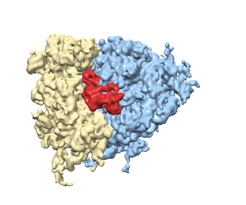 emd_8806.png emd_8806.png | 141.7 KB | ||
| アーカイブディレクトリ |  http://ftp.pdbj.org/pub/emdb/structures/EMD-8806ftp://ftp.pdbj.org/pub/emdb/structures/EMD-8806 http://ftp.pdbj.org/pub/emdb/structures/EMD-8806ftp://ftp.pdbj.org/pub/emdb/structures/EMD-8806 | HTTPS FTP |
-関連構造データ

-リンク
| EMDBのページ | EMDB (EBI/PDBe) / EMDataResource |
|---|---|
| 「今月の分子」の関連する項目 |

-マップ
| ファイル | ダウンロード / ファイル: emd_8806.map.gz / 形式: CCP4 / 大きさ: 144.7 MB / タイプ: IMAGE STORED AS FLOATING POINT NUMBER (4 BYTES) | ||||||||||||||||||||||||||||||||||||||||||||||||||||||||||||||||||||
|---|---|---|---|---|---|---|---|---|---|---|---|---|---|---|---|---|---|---|---|---|---|---|---|---|---|---|---|---|---|---|---|---|---|---|---|---|---|---|---|---|---|---|---|---|---|---|---|---|---|---|---|---|---|---|---|---|---|---|---|---|---|---|---|---|---|---|---|---|---|
| 注釈 | Rabbit 80S ribosome derived from EMPIAR-10064 using emClarity. Mid-rotated/swiveled 40S with eeF2 | ||||||||||||||||||||||||||||||||||||||||||||||||||||||||||||||||||||
| ボクセルのサイズ | X=Y=Z: 2.62 Å | ||||||||||||||||||||||||||||||||||||||||||||||||||||||||||||||||||||
| 密度 |
| ||||||||||||||||||||||||||||||||||||||||||||||||||||||||||||||||||||
| 対称性 | 空間群: 1 | ||||||||||||||||||||||||||||||||||||||||||||||||||||||||||||||||||||
| 詳細 | EMDB XML:
CCP4マップ ヘッダ情報:
| ||||||||||||||||||||||||||||||||||||||||||||||||||||||||||||||||||||
-添付データ
- 試料の構成要素
-全体 : 80s ribsome from Oryctolagus cuniculus
| 全体 | 名称: 80s ribsome from Oryctolagus cuniculus |
|---|---|
| 要素 |
|
-超分子 #1: 80s ribsome from Oryctolagus cuniculus
| 超分子 | 名称: 80s ribsome from Oryctolagus cuniculus / タイプ: complex / ID: 1 / 親要素: 0 |
|---|---|
| 由来(天然) | 生物種: Oryctolagus cuniculus (ウサギ) / 組織: Reticulocyte |
-実験情報
-構造解析
| 手法 | クライオ電子顕微鏡法 |
|---|---|
 解析 解析 | サブトモグラム平均法 |
| 試料の集合状態 | particle |
-試料調製
| 緩衝液 | pH: 7.6 構成要素:
| ||||||||||||
|---|---|---|---|---|---|---|---|---|---|---|---|---|---|
| グリッド | モデル: Quantifoil R2/1 / 材質: COPPER / 支持フィルム - 材質: CARBON / 支持フィルム - トポロジー: HOLEY / 前処理 - タイプ: PLASMA CLEANING | ||||||||||||
| 凍結 | 凍結剤: ETHANE / チャンバー内湿度: 60 % / チャンバー内温度: 295 K / 装置: FEI VITROBOT MARK IV |
- 電子顕微鏡法
| 顕微鏡 | FEI TITAN KRIOS |
|---|---|
| 電子線 | 加速電圧: 300 kV / 電子線源: FIELD EMISSION GUN |
| 電子光学系 | 照射モード: SPOT SCAN / 撮影モード: BRIGHT FIELDBright-field microscopy / Cs: 2.7 mm / 最大 デフォーカス(公称値): 3.7 µm / 最小 デフォーカス(公称値): 2.4 µm |
| 詳細 | Data collection 20 --> -60, 22 --> 60 degrees |
| 撮影 | フィルム・検出器のモデル: GATAN K2 SUMMIT (4k x 4k) 検出モード: INTEGRATING / デジタル化 - サイズ - 横: 3838 pixel / デジタル化 - サイズ - 縦: 3710 pixel / 平均電子線量: 1.5 e/Å2 |
| 実験機器 |  モデル: Titan Krios / 画像提供: FEI Company |
-画像解析
| 抽出 | トモグラム数: 4 / 使用した粒子像数: 3090 |
|---|---|
| CTF補正 | ソフトウェア - 名称: emClarity (ver. 1.0) ソフトウェア - 詳細: Included astigmatism and per-tilt CTF determination 詳細: Phases corrected on the projections by multiplying by the CTF in tiles compensating for the defocus gradient in tilted images. |
| 最終 3次元分類 | クラス数: 5 / 平均メンバー数/クラス: 500 / ソフトウェア - 名称: emClarity (ver. 1.0) 詳細: 3,090 total subtomograms were assigned to 5 separate classes using multi-scale PCA with full-3dCTF compensated estimators. 1.0,1.6,2.4,5.4 nm were the length-scales used for the msPCA. |
| 最終 角度割当 | タイプ: OTHER / ソフトウェア - 名称: emClarity (ver. 1.0) 詳細: 3D refinement with two half-sets fully separated after template matching, at which point the FSC 0.5 was 3.6 nanometers. Reference and subtomograms filtered dynamically according to the FSC ...詳細: 3D refinement with two half-sets fully separated after template matching, at which point the FSC 0.5 was 3.6 nanometers. Reference and subtomograms filtered dynamically according to the FSC at each cycle using the FOM approach. |
| 最終 再構成 | 使用したクラス数: 1 / 想定した対称性 - 点群: C1 (非対称) / アルゴリズム: BACK PROJECTION / 解像度のタイプ: BY AUTHOR / 解像度: 11.0 Å / 解像度の算出法: FSC 0.143 CUT-OFF / ソフトウェア - 名称: emClarity (ver. 1.0) ソフトウェア - 詳細: adapted single-particle wiener filter 詳細: Independent beyond 3.6 nm as determined by the FSC at the time of dividing the half-sets 使用したサブトモグラム数: 546 |
| FSC曲線 (解像度の算出) | 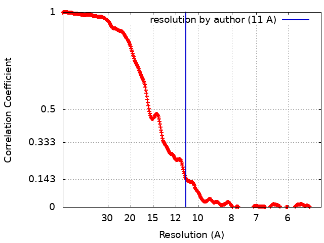 |
